9
MRI feet - accurate diagnosis of diseases and injuries
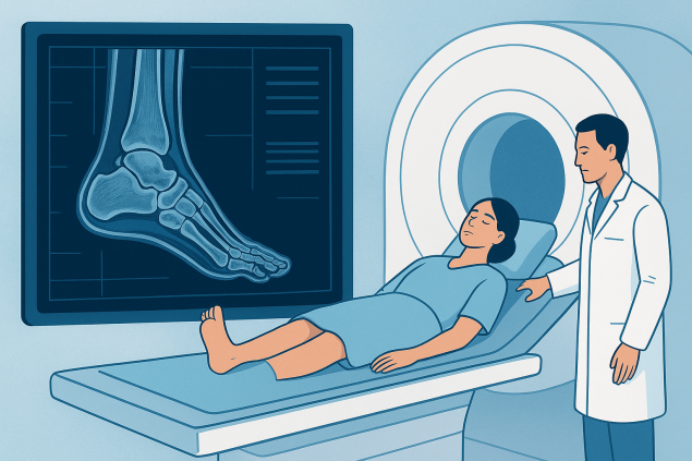
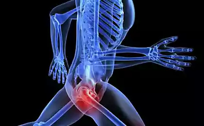
Modern medicine offers many methods of research, but when it comes to complex structures such as joints, ligaments and soft tissues, the most informative is the mRI of the foot. This procedure allows you to accurately identify damage to bones, cartilage, tendons and blood vessels, which makes it indispensable for injuries, chronic pain or suspected inflammatory processes. Unlike x-rays or ultrasound, magnetic resonance imaging gives a layer-by-layer image of tissues, allowing the doctor to consider the smallest changes that cannot be detected by other methods.
MRI of the foot is carried out painlessly and safely, as it does not use ionizing radiation. The patient is placed in a tomograph machine, where detailed images are created using a magnetic field and radio waves. The procedure takes 20 to 40 minutes, and the results can be used both for initial diagnosis and to monitor the effectiveness of treatment.

When is prescribed MRI feet and what are the advantages of the method
This study is recommended for the following conditions:
- foot injuries (fractures, dislocations, damage to ligaments and tendons);
- chronic pain of unknown origin;
- suspected arthritis, arthrosis or gout;
- inflammatory diseases of soft tissues;
- neoplasms, cysts, tumors;
- assessment of the condition after surgery;
- Diagnosis of complications in diabetes (for example, diabetic foot).
The advantages of magnetic resonance imaging are obvious:
- high accuracy of the study;
- the ability to see not only bones, but also ligaments, muscles, vessels;
- absence of pain and harmful radiation;
- identification of pathologies in the early stages;
- the ability to monitor the dynamics of treatment.
Unlike radiography, which only shows bones, MRI gives a complete picture of the state of all tissues. This is especially important when it comes to sports injuries or chronic diseases, when the most accurate diagnosis is required.
How is the MRI procedure of the foot
The patient is located on a special table, the leg is fixed to exclude movements. It is important to remain still to get clear pictures. In some cases, a contrast agent is used, which is injected intravenously for more detailed imaging of blood vessels and soft tissues. Contrast is absolutely safe and quickly excreted from the body.
During the scan, the patient hears the characteristic clicks of the device, so headphones or earplugs can be used. After the study is completed, the results are processed by a radiologist, and the patient receives a detailed conclusion.
Why you should choose an MRI
MRI foot is one of the most informative and safe diagnostic methods. It helps to identify problems in time, start treatment and prevent complications. Leading clinics use the latest generation of tomographs, which provide maximum accuracy and comfort for the patient.
This study is recommended not only in the presence of symptoms, but also as a preventive measure for athletes, people with chronic joint diseases and patients after injuries.
MRI of the foot has become the gold standard of diagnosis, which allows the doctor to make an accurate diagnosis and choose the optimal treatment. Thanks to this method, patients are able to maintain health, mobility and activity for many years.


Plastic containers and shelving: the basis of effective storage
7 things that prevent you from taking better care of yourself