1409
The bones in the legs
The narrow, graceful female feet in a beautiful dress shoes - object of admiration for the men and the theme of poetry enthusiastic ... Alas, for some women it is only a dream. In reality - buy shoes 1-2 sizes larger (because "a" does not fit the size of the foot), and a painful, enlarged in diameter stop. Blame the deformation in the first metatarsus-phalanx joints - the so-called pits on his feet.
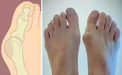
Almost always there is another component - a deviation of the big toe outwards (medical name - hallux valgus), and the first metatarsal - medially (Figure 2). It was the head of the first metatarsal bone forms a protrusion at the base of the first finger - bone. Changing the width of the foot. As a result of compression of the shoes of soft tissues in the bones may be inflammation of the bursa, while the symptoms of redness and swelling joins, this place becomes painful. On top of pits on the feet are small. Reject the first finger moves the head of the first metatarsal bone to the edge of the foot, pressure from shoes on her intensified. Over time, the edge of the head bone growths appear, and we are seeing an increase in seed. First metatarsal bone is not only deflected to the side, and raised above the support. While walking load on it is reduced, but increasingly beginning to be loaded 2-3 metatarsals. On the soles, under the heads of these bones are formed corns, often painful. Disturbed muscle balance and imbalance leads to a shift to the rear 2-3-4 fingers to form a hammer deformation. Calluses on the protruding parts of the deformed foot deliver a lot of trouble to her mistress.
According to statistics, the bones in the feet to 35-40 years formed one in three of the fair sex. What is this pathology? The name "bone on his feet" does not fully reflect the essence of the problem. Upon closer examination, even in the initial stages in addition to the "bones" of the inner surface of the foot, you can see the deviation of the first toe outwards (hallux valgus), and the first metatarsal bone medially. Increasing the width of the foot, there are difficulties in the selection of shoes. At the beginning of "pits" on the feet have a small size. At this stage, it is mainly composed of an offset head of the first metatarsal.
As a result of compression of the shoes of soft tissues in the "pits" may be inflammation of the bursa, there are pain, swelling and redness. The head of the first metatarsal bone is subjected to the same pressure on the edge of its growths appear. As a result - "Bone" further increases in size.
In more severe cases appear hammer deformation of the second and third fingers, often develops deforming arthrosis of the first metatarsus-phalanx joint. First metatarsal bone is not only deflected to the side, and raised above the support. While walking load on it is reduced, but increasingly beginning to be loaded 2-3 metatarsals. On the soles, under the heads of these bones are formed corns, often painful (Figure 1).
Components deformity
Fig.1 "Stone" on the leg - a multi-component deformation
In addition exostosis (actually "bones") is usually present deviation of the first toe outwards (hallux valgus). Due to the lowering of 2-3 metatarsals soles are formed corns (shown in red), develops hammer deformation of 2-3 fingers and toes on their back surface (marked in blue).
Depending on the severity, severity of symptoms deformation specialists distinguish three of its extent (Figure 2).
The degree of deformation in the foot bones in the legs
Fig.2 "Stone" on the foot develops gradually. On the left - a moderate strain, then - moderate right - expressed
As you can see, "bones" on the leg - a complex multi-component deformation of the forefoot. Conventionally, the line between cosmetic deformity and disease takes place where there is pain when walking and impaired locomotor function.
At the beginning of the disease can give the effect of conservative treatment. Stabilization of the first metatarsal bone with the help of individual insoles, the use of special, the first finger abductor, tires, interdigital pads, braces, exercises aimed at restoring muscle imbalance and eliminate contractures formed - in the complex all this can stop the progression of the disease. If conservative methods have no effect on the legs and bones continue to grow, or strain already expressed, surgery can help.
Medicine does not stand still, every year we can better understand the causes of diseases, development of new techniques to remove the deformation based on the latest achievements in traumatology and orthopedics. Modern anesthetic warrants pain during surgery. Preoperative planning and carefully crafted postoperative provide full rehabilitation of our patients.
Remove bones of the feet in the center of plastic surgery
Surgical treatment of bones on their feet, doctors involved in the time of Hippocrates and Antillus. Typically, 90% of all techniques intended to change the position of the first metatarsal. However, until recently, to keep it in the correct position, used unreliable locks, had to further impose a plaster cast on 1, 5-2 months. And the longer the fixed limb in a cast, the more difficult the subsequent rehabilitation.
Currently, the treatment of the above diseases increasingly using combined types of operations on the bones and soft tissues of the foot. The main disadvantage of such treatment is the necessity of immobilization plaster cast followed by prolonged rehabilitation.
In our clinic we use modern techniques to help the patient in a state after surgery do without gypsum. We believe (and many surgeons will agree) that every patient needs a differentiated approach. The degree of deformation, the presence of concomitant flat feet, or vice versa, hollow feet, changes in the first metatarsophalangeal joint, metacarpophalangeal, age - these and other factors must be considered when choosing treatment. In one case, we can restrict only the intervention of the soft tissues in the other - required the combined surgery on the bones of the forefoot combined with the operation in the muscles and tendons in the third - the expanded intervention on the bones.
In our arsenal there are several surgical techniques that allow you to achieve the desired aesthetic and functional results. In this rehabilitation period it is considerably easier, compared with other conventional methods.
Advantages of our methods:
the possibility of simultaneous correction of all components of the deformation of the forefoot;
Small traumatic intervention;
if you have to cross the bone (osteotomy do), we use it such a form that provides the greatest stability. Bone heals faster and the patient is able to walk to start from the very first days after the operation;
We try not to leave foreign materials in the area of intervention, either to reduce their number to a minimum;
do not use a plaster cast;
This approach significantly reduces the time of treatment and rehabilitation;
the operation is performed under local anesthesia or wires.
After the operation, we impose normal bandage. On the same day, or the next allow the load on the heel. At the end of the treatment you can wear normal shoes, including, and heels.
Read more about the procedures of pitting on the legs
Correcting the position of the first metatarsal osteotomy through (the intersection)
From a small incision in the "pits" to dissect the soft tissue. Remove excessively-grown soft tissue and the part of the "bumps" on the foot, which is not involved in normal operation of the joint. Special thin-bladed saw sagittal cross the first metatarsal bone. And the plane of intersection is located in the space so that there is no tendency halves bone spontaneously move during walking, and hence the risk of nonunion minimized. Sometimes it is necessary to complement osteotomy also manipulation of soft tissues - ligaments release that are holding the first finger in the retracted position. The intersection of bone laid in the correct position (the head of the first metatarsal bone is getting closer to the second and below). Locks the two titanium screws, screw heads are immersed so that they did not act in the future under the skin. The position with the finger is corrected. The foot becomes.
Intervention of soft tissue
Two small incisions are accessing the zone of intervention, carried out manipulation of the tendons and muscles, move the first finger joint capsule, correct finger position, unload the joint. From the capsule, tendons, ligaments, cuts out and form a new anatomical structures with which bring together metatarsals, eliminate the offset sesamoid bones. Externally stop becomes normal anatomical shape.
After the operation, we impose normal bandage, the next day the patient to allow walking footwear. In such orthopedic sandals have to go 5 weeks. After healing metatarsal swelling has subsided and the patient returns to normal shoes, among other things, on the heel.
clinicaveka.ru/