496
New infrared laser finds the cancerous cells
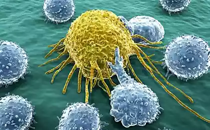
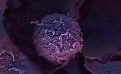
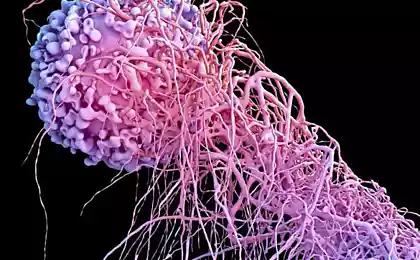
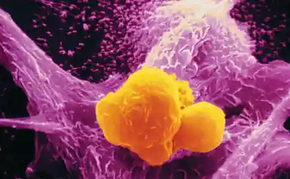
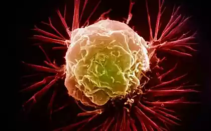
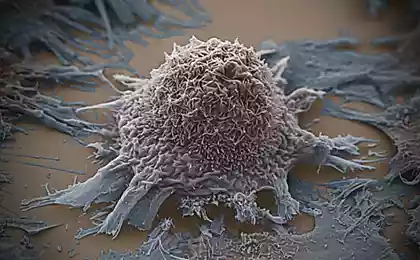
The method is based on a feature of cancer cells, which is associated with the location in them of mitochondria — organelles responsible in the cell for energy production. One cell may be a couple of hundred or even thousands of mitochondria. They usually concentrate around the nucleus of the cell and carefully avoid proximity to the cell membrane. But in cancer cells the mitochondria are almost evenly distributed throughout the volume of the cytoplasm.
This fact is know for a long time, but only last year in Sandia lab could use it to identify cancer cells. The size of mitochondria amounted to near 800 nm. Therefore, 90 to 95% of laser radiation with a wavelength of 800 nm is scattered on them, and through other cell components such radiation passes almost freely. The nature of the observed scattering and fluorescence strongly depends on how the mitochondria are distributed within cells.
Short pulse semiconductor laser is focused on a single cell and its scattering on mitochondria instantly becomes clear, healthy cell or not. However, for the analysis of real clinical samples, laser sorting system, it was necessary to include a special conveyor, which delivers the cells singly.
Source: /users/413

This fact is know for a long time, but only last year in Sandia lab could use it to identify cancer cells. The size of mitochondria amounted to near 800 nm. Therefore, 90 to 95% of laser radiation with a wavelength of 800 nm is scattered on them, and through other cell components such radiation passes almost freely. The nature of the observed scattering and fluorescence strongly depends on how the mitochondria are distributed within cells.
Short pulse semiconductor laser is focused on a single cell and its scattering on mitochondria instantly becomes clear, healthy cell or not. However, for the analysis of real clinical samples, laser sorting system, it was necessary to include a special conveyor, which delivers the cells singly.

Source: /users/413
Bike with electric motor: a worthy replacement for compact cars
Tesla promises driverless cars within three years
