1185
Beauty under the microscope
An inspiring sight fantastic 3D images of tissue and surrounding mira.Elektronny microscope - is an amazing tool that allows you to see the smallest particles, present and understand what can not be seen with the naked eye. Unlike optical microscope, electron microscope produces images of objects with the greatest increase to 10 6 times. And a new world opens up to people - the volume and color.
Scanning microscope works on the principle of scanning television thin beam of electrons on the surface of the sample, allowing you to get an inspirational 3D images of tissue. Or, for example, a snowflake and the human louse on the hair, and the muzzle of a mosquito, shark skin or fat cells of the human body.
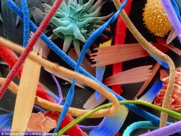
Household dust
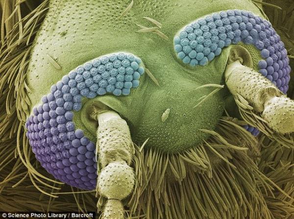
Komar
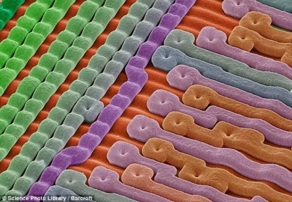
The surface of a silicon microchip
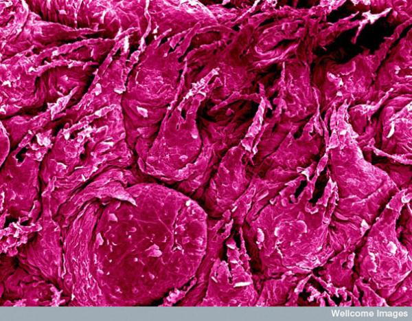
The surface of the tongue
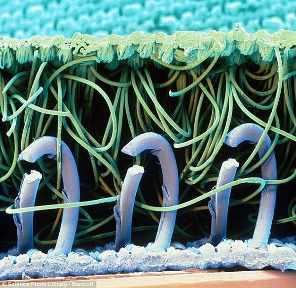
Velcro
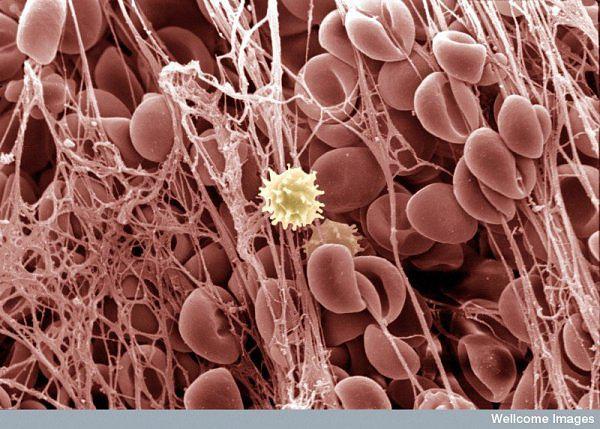
A blood clot
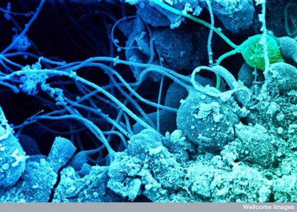
Sperm develop in the testes
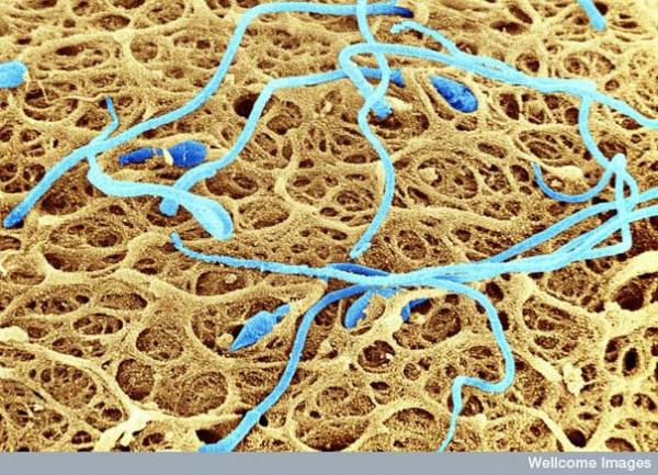
Sperm on the surface of the egg
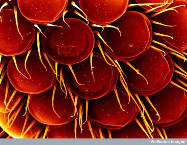
Eye gnats
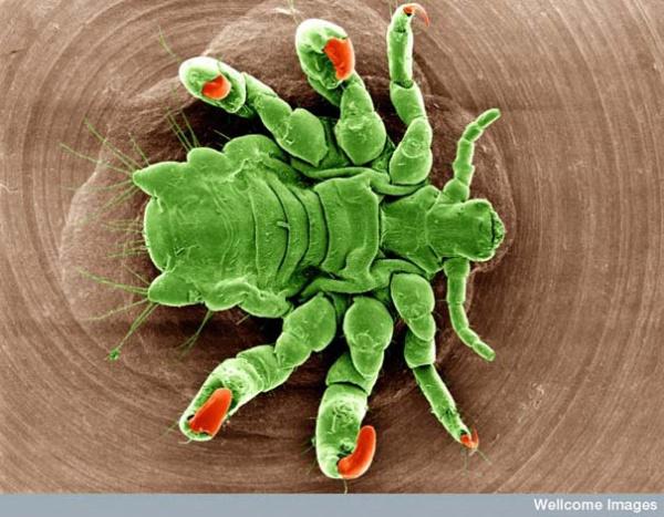
Pubic lice
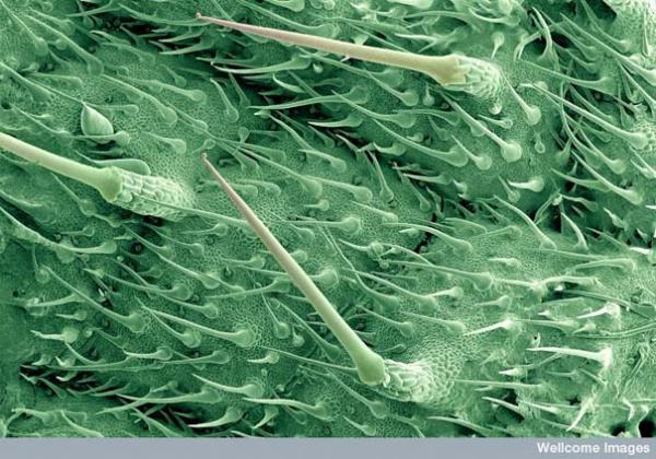
Burning hair nettle leaves
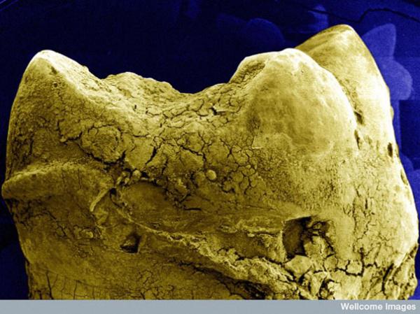
The surface of the tooth
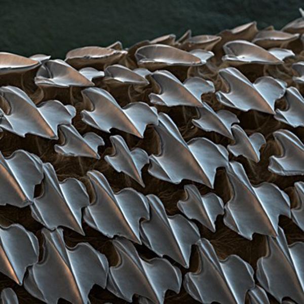
Sharkskin
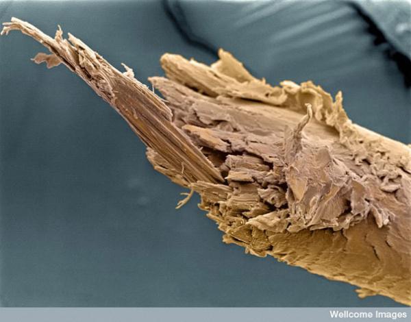
The cut of a human hair
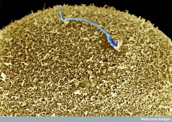
Sperm on the surface of the egg
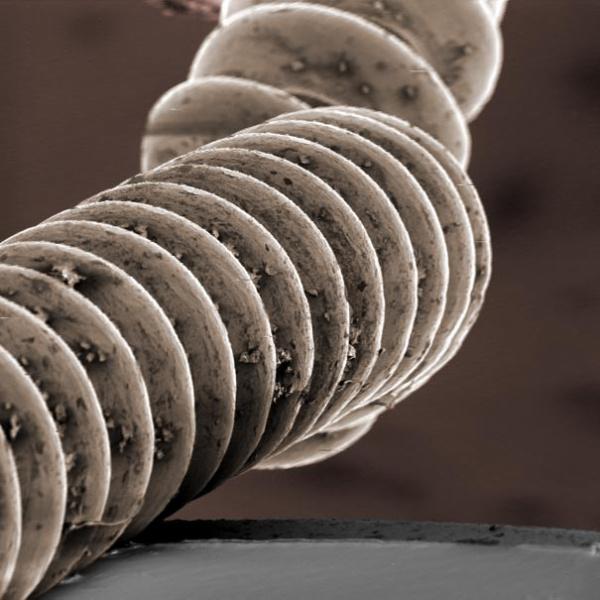
String electric
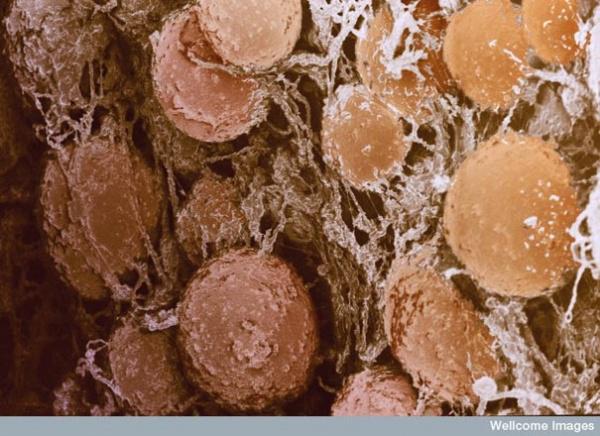
Fat cells
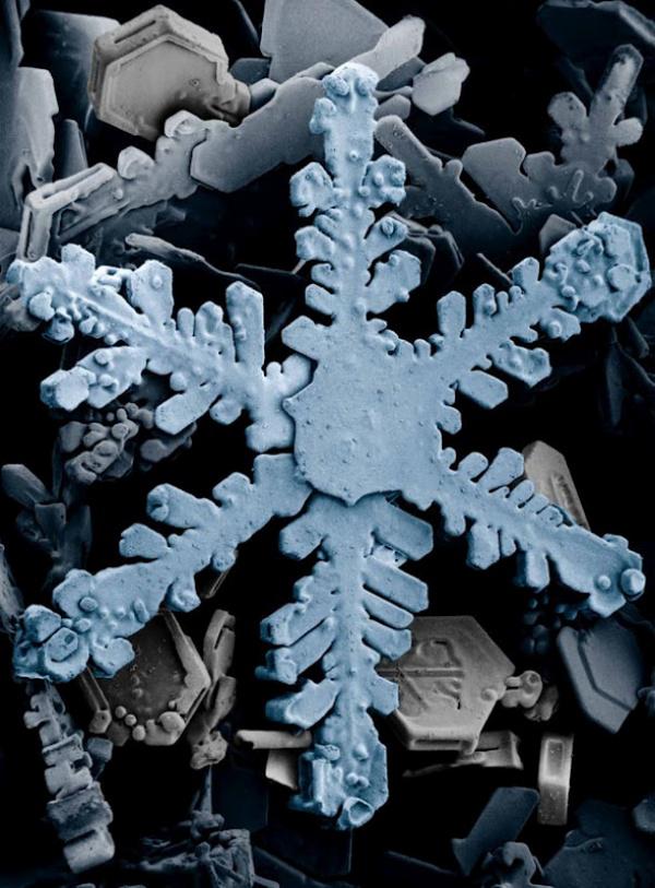
Snow
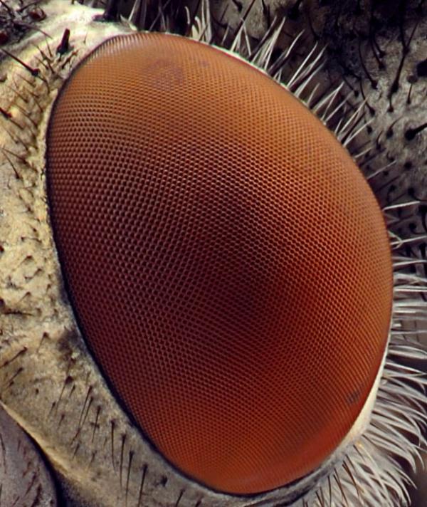
Eye fly
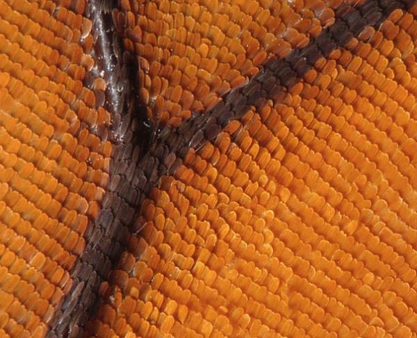
Butterfly wing
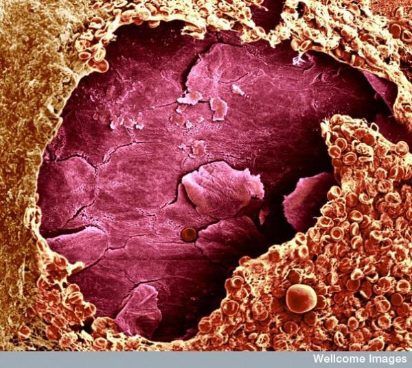
Tighten the wound
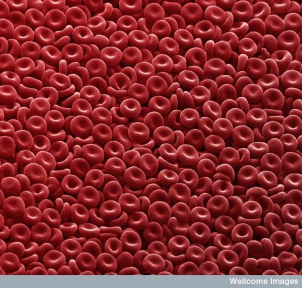
Red blood cells
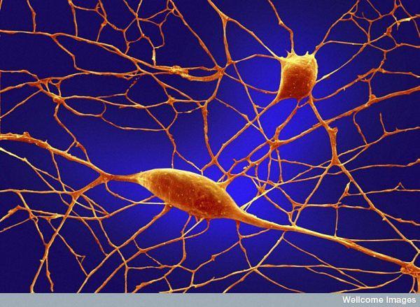
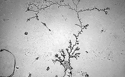
Neurons in the brain
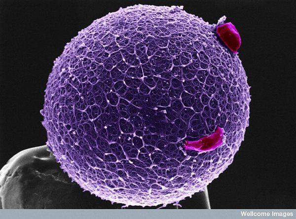
The egg
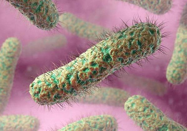
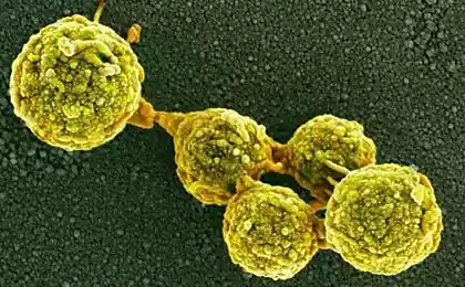
Rod-shaped bacteria ciliated
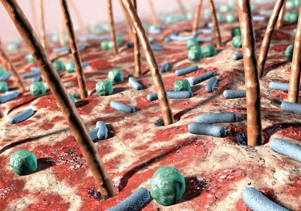
The bacteria on human skin
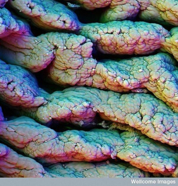
The villi of the small intestine
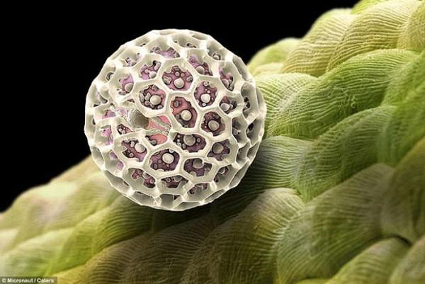
Pollen
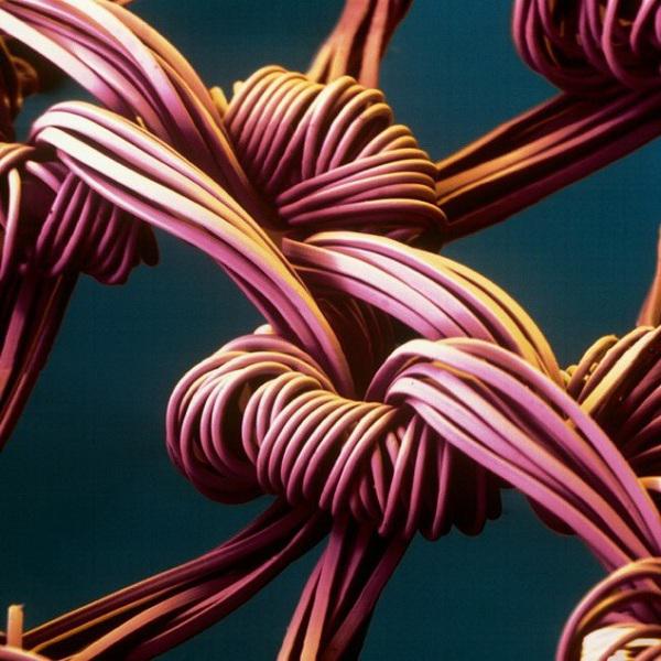
Nylon stockings
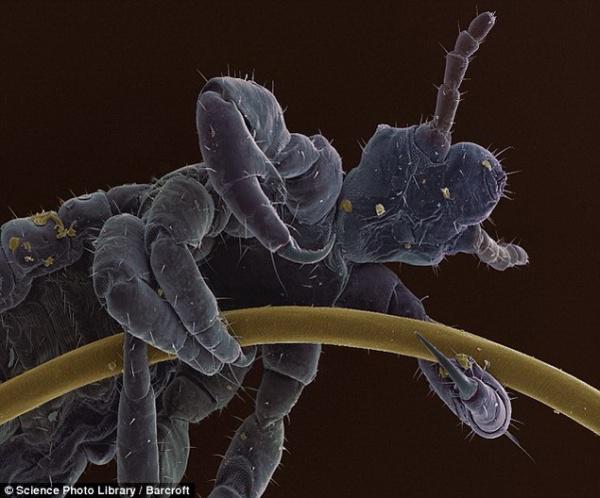
The human louse on the hair
See also:
Fabulous macro photography of insects
via www.adme.ru/vdohnovenie-919705/skazochnye-makrofotografii-nasekomyh-340705/
Scanning microscope works on the principle of scanning television thin beam of electrons on the surface of the sample, allowing you to get an inspirational 3D images of tissue. Or, for example, a snowflake and the human louse on the hair, and the muzzle of a mosquito, shark skin or fat cells of the human body.

Household dust

Komar

The surface of a silicon microchip

The surface of the tongue

Velcro

A blood clot

Sperm develop in the testes

Sperm on the surface of the egg

Eye gnats

Pubic lice

Burning hair nettle leaves

The surface of the tooth

Sharkskin

The cut of a human hair

Sperm on the surface of the egg

String electric

Fat cells

Snow

Eye fly

Butterfly wing

Tighten the wound

Red blood cells

Neurons in the brain

The egg

Rod-shaped bacteria ciliated

The bacteria on human skin

The villi of the small intestine

Pollen

Nylon stockings

The human louse on the hair
See also:
Fabulous macro photography of insects
via www.adme.ru/vdohnovenie-919705/skazochnye-makrofotografii-nasekomyh-340705/


