787
Macromir
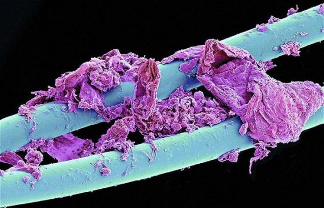
1. Use dental floss at high magnification looks awful. Better not go into details of what the pink lumps on a blue nylon. How good that is in the range of conventional fluorescent light, it is, in principle, impossible to see.
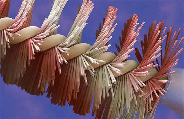
2. Unused brush mascara looks much more attractive even under an electron microscope.
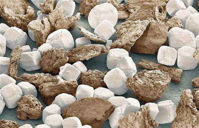
3. These colored stones are actually a grain of salt and black pepper from a jar with spices.
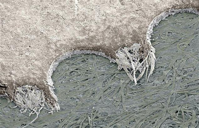
4. It is difficult to believe that glossy stamp actually has such a loose and fibrous structure. The picture shows the torn edge of a postage stamp.
5. Another monster from the bathroom - Use cotton swab to clean the ears.
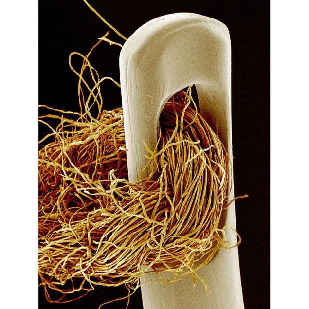
6. The naked human eye can distinguish objects, the size of which is not less than 0 176 mm. The best optical microscopes are limited in their work wavelength of visible light, and therefore, they can not provide details of observation which is less than 0, 1-0, 2 microns. Electron microscope scans the sample with an electron beam and allows us to consider the details already nanometer. However, the sample for examination under the electron microscope is necessary to prepare a special way. The picture shows the eye of a needle with thread vdet.
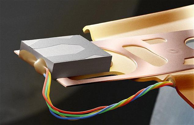
7. Scientific photographer Steve Gshmayssner of Bedfordshire after retirement continue to enjoy access to the scanning electron microscope, and can make interesting pictures. This picture shows a part of the computer chip.
8. These logs are actually hair, cut electric shaver blades.
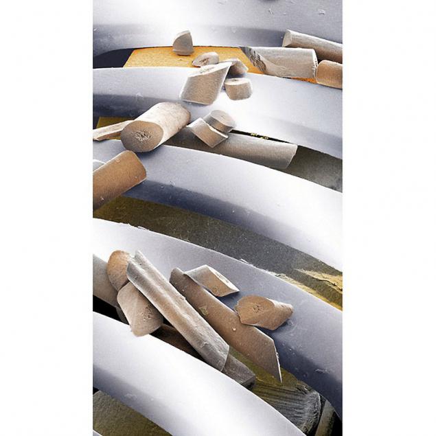
9. The cost of a scanning electron microscope is from 150 000 to 500 000 pounds. Of course, such a device can only afford a large scientific or industrial laboratory. Therefore Gshmayssner very pleased that after retirement, he has the opportunity to "play" with such a serious tool. The photos come out really very interesting. The picture shows the structure of a guitar string.
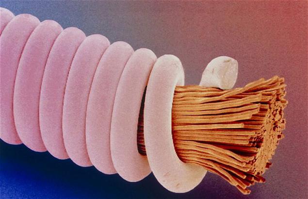
10. Normal zip fastener.
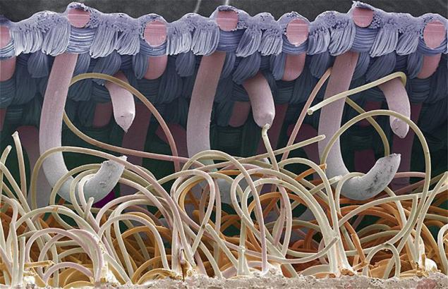
11. pyrophoric flint from the ignition device of a conventional lighter.
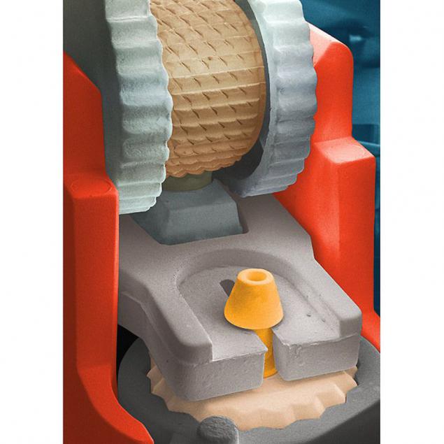
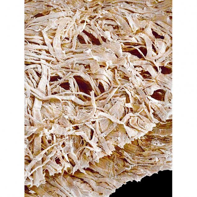
12. The loose structure of the toilet paper.
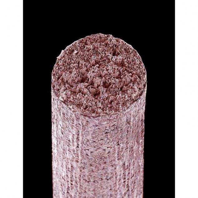
13. Writing a simple rod in graphite pencil.
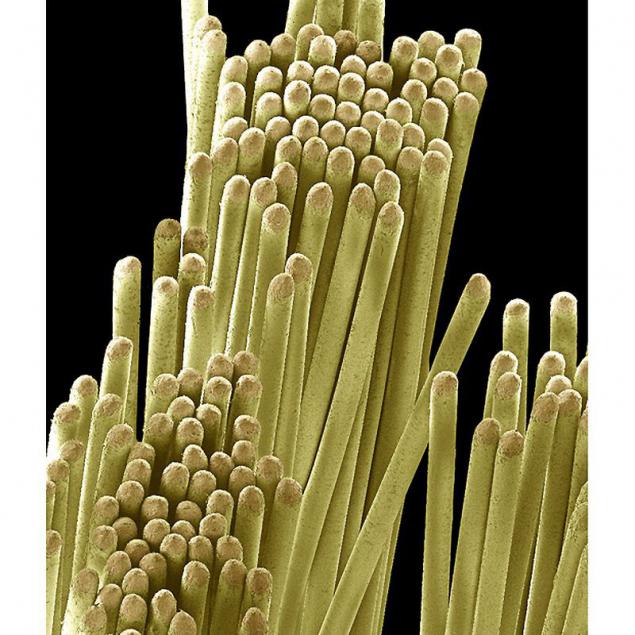
14. The structure of the toothbrush bristles.
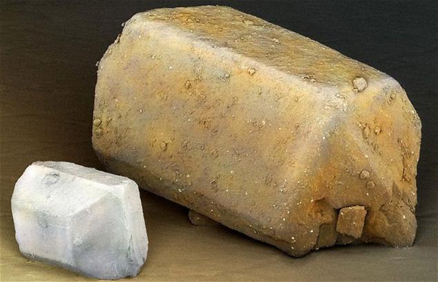
15. Crystals of refined sugar and brown sugar.
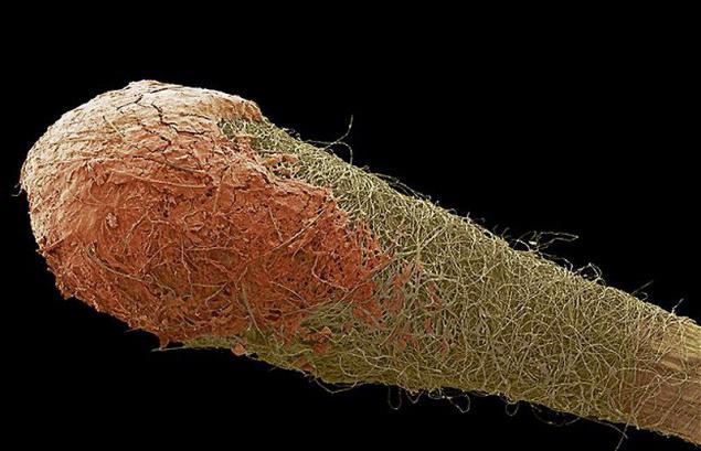
16. And looks match head under a microscope.

2. Unused brush mascara looks much more attractive even under an electron microscope.

3. These colored stones are actually a grain of salt and black pepper from a jar with spices.

4. It is difficult to believe that glossy stamp actually has such a loose and fibrous structure. The picture shows the torn edge of a postage stamp.

5. Another monster from the bathroom - Use cotton swab to clean the ears.

6. The naked human eye can distinguish objects, the size of which is not less than 0 176 mm. The best optical microscopes are limited in their work wavelength of visible light, and therefore, they can not provide details of observation which is less than 0, 1-0, 2 microns. Electron microscope scans the sample with an electron beam and allows us to consider the details already nanometer. However, the sample for examination under the electron microscope is necessary to prepare a special way. The picture shows the eye of a needle with thread vdet.

7. Scientific photographer Steve Gshmayssner of Bedfordshire after retirement continue to enjoy access to the scanning electron microscope, and can make interesting pictures. This picture shows a part of the computer chip.

8. These logs are actually hair, cut electric shaver blades.

9. The cost of a scanning electron microscope is from 150 000 to 500 000 pounds. Of course, such a device can only afford a large scientific or industrial laboratory. Therefore Gshmayssner very pleased that after retirement, he has the opportunity to "play" with such a serious tool. The photos come out really very interesting. The picture shows the structure of a guitar string.

10. Normal zip fastener.

11. pyrophoric flint from the ignition device of a conventional lighter.

12. The loose structure of the toilet paper.

13. Writing a simple rod in graphite pencil.

14. The structure of the toothbrush bristles.

15. Crystals of refined sugar and brown sugar.

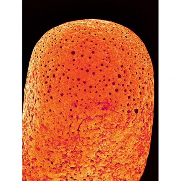
16. And looks match head under a microscope.

