483
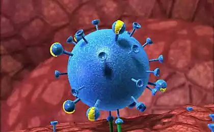
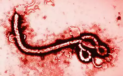
Unique 3D-video, the virus struggles to infect cells
Pristavuchie mikroorganizmNovy imaging technique demonstrates the activity of the virus and how it is introduced into the cell. The video shows a small ball, which randomly tossing until it hits the cell. Then, it bounces and moves along a surface, either takes off again, or insidiously slips into the cell. At first glance it seems that the virus behaves erratically, but in fact it is working mechanism oiled.
The virus in the video - it is an artificial microscopic beads of polystyrene, studded with protein segments, known as Tat-peptides - is derived from HIV-1, they help to find the particle cell. The ball is equipped with quantum dots (semiconductor or bits), emits light that helps the camera to find and track it.
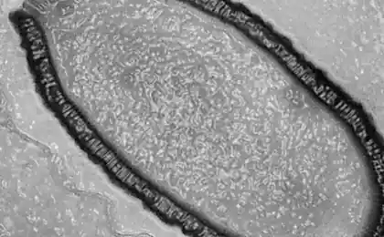
Movement of the virus particle and high granularity circuits cells were recorded using a technique called laser scanning microscopy: a chamber holding the frame in the particle, and the other is directed to the cell. As a result, 3D-visualization in real time is a work of two cameras. Before this method is detailed image could only give micron microscope, but the images in this case are two-dimensional.
The possibility thus follow the movements of the particles has allowed scientists to observe the very small structures with a precision of about ten nanometers, which used to be possible to see only with an electron microscope. A nanometer is one billionth of a meter, and is about 1000 times thinner than a human hair.
Ultimately, this research will help scientists to deal with different infections, as well as contribute to the development of nanotechnology and medical microscopic machines.
via factroom.ru
The virus in the video - it is an artificial microscopic beads of polystyrene, studded with protein segments, known as Tat-peptides - is derived from HIV-1, they help to find the particle cell. The ball is equipped with quantum dots (semiconductor or bits), emits light that helps the camera to find and track it.
Movement of the virus particle and high granularity circuits cells were recorded using a technique called laser scanning microscopy: a chamber holding the frame in the particle, and the other is directed to the cell. As a result, 3D-visualization in real time is a work of two cameras. Before this method is detailed image could only give micron microscope, but the images in this case are two-dimensional.
The possibility thus follow the movements of the particles has allowed scientists to observe the very small structures with a precision of about ten nanometers, which used to be possible to see only with an electron microscope. A nanometer is one billionth of a meter, and is about 1000 times thinner than a human hair.
Ultimately, this research will help scientists to deal with different infections, as well as contribute to the development of nanotechnology and medical microscopic machines.
via factroom.ru