695
The oral cavity and its contents
What you see in these photos seem to rare plants and exotic landscapes, but in fact - is ... bacteria that nestled on your teeth, as well as other micro-organisms living on the gums or on the toothbrush.
These macro photography were made with the help of a microscope, which scans a sample by a focused electron beam. After that the pictures were painted digitally or manually so that you can distinguish between the individual elements. These pictures belong to the Scientific darkroom in London and are used for research and educational purposes. They show us the consequences of improper oral hygiene.
22 photos via telegraph.co.uk
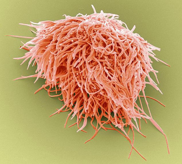
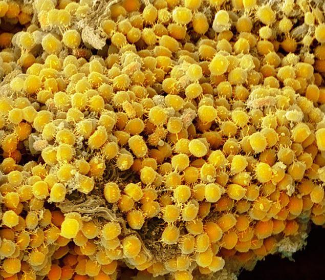
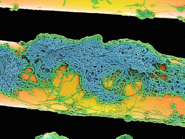
2. Plaque (magnification of 400 times, the width of the picture - 10 cm) - is a biofilm formed by bacteria colonizing that attempt to join the tooth surface.
3. And this is the same plaque at a magnification of 10 000 times. The width remains unchanged.
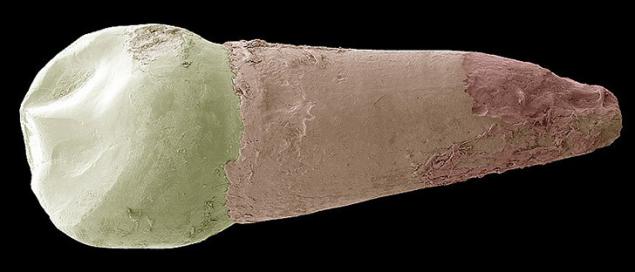
4. Milk tooth. Most human teeth formed from the dentine - substances shroud cavity which are soft connective tissue, blood vessels and nerves. Then the crown of the tooth enamel covers (pictured above white) - harder and mineralized material that protects the dentin of the acids in the mouth. At the root of the tooth dentin is protected by a substance called cement (pink), which serves as a means by which the periodontal ligament can be attached to the tooth stability
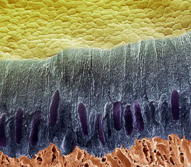
5. This shows amelogenic layer of cells (blue), the surface of the teeth (yellow) and dentin (red). Loss of enamel or dentin exposes cement - porous material with microscopic channels called dentinal tubules which connect the pulp, which leads to tooth sensitivity.
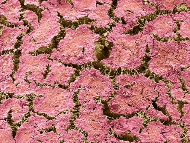
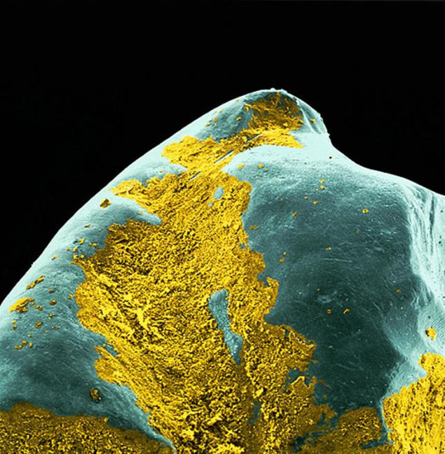
6. The yellow shows the plaque on the tooth surface. Acid is a waste generated during the digestive process of the bacteria. She deminiralizuet tooth, creating cavities to be filled or that can lead to tooth loss.
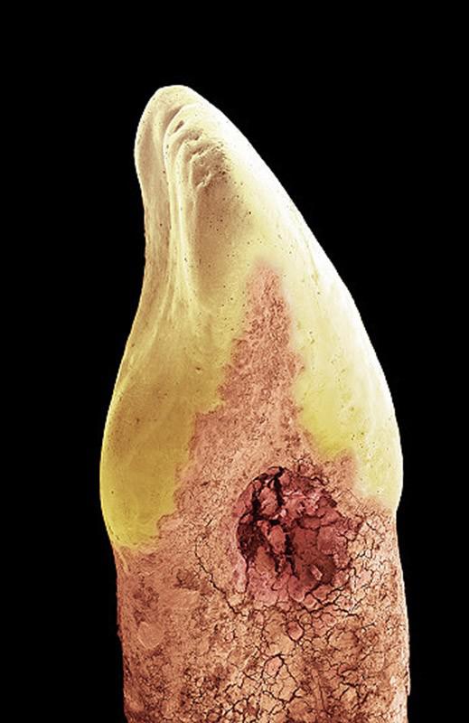
7. The human tool with a hollow or loss of minerals caused by bacterial acid "garbage." In this case, the cavities formed on the side of the tooth (between two teeth) and at the gum line (between the crown - yellow - and the root), possibly due to lack of flossing or improper use. British scientists have found that one in three adult susceptible to caries, as a study conducted in the 5-year children in 2012 showed that every fourth of them have some degree of dental caries.
8. We hope this photo will convince you to brush your teeth every day thread ... Yellow - are bacteria on the gums. Plaque accumulation may cause damage to the gums, such as gingivitis or periodontitis.
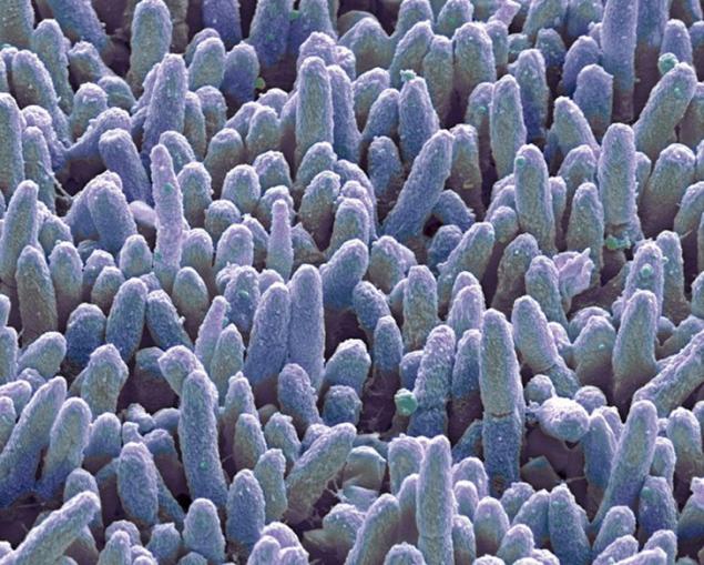
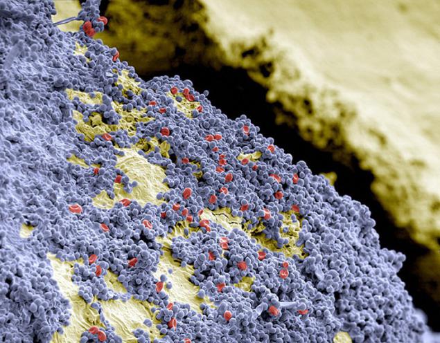
9. The surface of human teeth (colored yellow) carpet spherical bacteria (colored blue) and blood vessels (colored red).
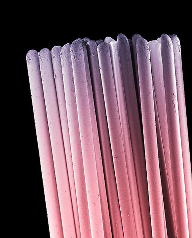
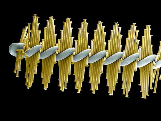
10. The bristles of the toothbrush. They erased and wear out over time, reducing the effectiveness of the toothbrush. Change toothbrush necessary, at least every 3-4 months. However, the wear may vary among individuals, depending on their habits brushing teeth. If necessary, the toothbrush should be changed first.
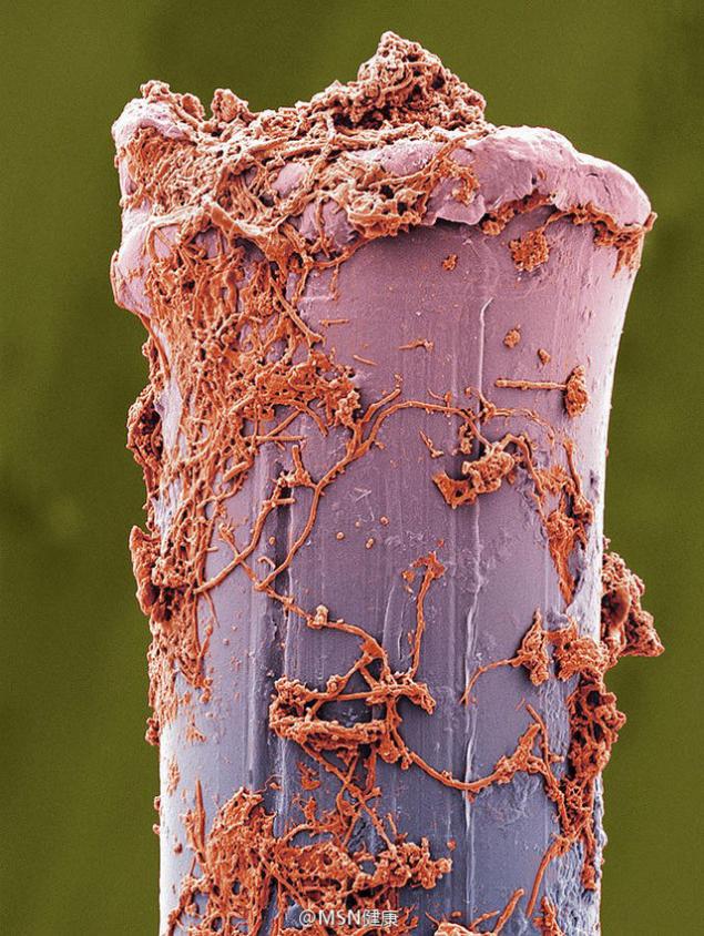
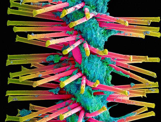
11. The bristles of the toothbrush, coated plaque. After brushing toothbrush should be thoroughly washed to remove all traces of toothpaste or pieces of plaque and bacteria. It should be dried by standing in the open air. The closed containers more moisture, which promotes the development of microorganisms.
12. Plaque used on the bristles of the toothbrush at a magnification of 750 times.
13. The interdental brushes (as in the photo) have small heads with bristles designed for cleaning between the teeth. Your dentist may advise you to use interdental brush, but it is not a substitute for dental floss.
14. A more detailed view of the interdental bristles used their brush covered with plaque.
These macro photography were made with the help of a microscope, which scans a sample by a focused electron beam. After that the pictures were painted digitally or manually so that you can distinguish between the individual elements. These pictures belong to the Scientific darkroom in London and are used for research and educational purposes. They show us the consequences of improper oral hygiene.
22 photos via telegraph.co.uk

2. Plaque (magnification of 400 times, the width of the picture - 10 cm) - is a biofilm formed by bacteria colonizing that attempt to join the tooth surface.

3. And this is the same plaque at a magnification of 10 000 times. The width remains unchanged.

4. Milk tooth. Most human teeth formed from the dentine - substances shroud cavity which are soft connective tissue, blood vessels and nerves. Then the crown of the tooth enamel covers (pictured above white) - harder and mineralized material that protects the dentin of the acids in the mouth. At the root of the tooth dentin is protected by a substance called cement (pink), which serves as a means by which the periodontal ligament can be attached to the tooth stability

5. This shows amelogenic layer of cells (blue), the surface of the teeth (yellow) and dentin (red). Loss of enamel or dentin exposes cement - porous material with microscopic channels called dentinal tubules which connect the pulp, which leads to tooth sensitivity.

6. The yellow shows the plaque on the tooth surface. Acid is a waste generated during the digestive process of the bacteria. She deminiralizuet tooth, creating cavities to be filled or that can lead to tooth loss.

7. The human tool with a hollow or loss of minerals caused by bacterial acid "garbage." In this case, the cavities formed on the side of the tooth (between two teeth) and at the gum line (between the crown - yellow - and the root), possibly due to lack of flossing or improper use. British scientists have found that one in three adult susceptible to caries, as a study conducted in the 5-year children in 2012 showed that every fourth of them have some degree of dental caries.

8. We hope this photo will convince you to brush your teeth every day thread ... Yellow - are bacteria on the gums. Plaque accumulation may cause damage to the gums, such as gingivitis or periodontitis.

9. The surface of human teeth (colored yellow) carpet spherical bacteria (colored blue) and blood vessels (colored red).

10. The bristles of the toothbrush. They erased and wear out over time, reducing the effectiveness of the toothbrush. Change toothbrush necessary, at least every 3-4 months. However, the wear may vary among individuals, depending on their habits brushing teeth. If necessary, the toothbrush should be changed first.

11. The bristles of the toothbrush, coated plaque. After brushing toothbrush should be thoroughly washed to remove all traces of toothpaste or pieces of plaque and bacteria. It should be dried by standing in the open air. The closed containers more moisture, which promotes the development of microorganisms.

12. Plaque used on the bristles of the toothbrush at a magnification of 750 times.

13. The interdental brushes (as in the photo) have small heads with bristles designed for cleaning between the teeth. Your dentist may advise you to use interdental brush, but it is not a substitute for dental floss.

14. A more detailed view of the interdental bristles used their brush covered with plaque.