2024
Small world in macro photo
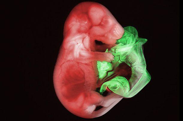
17-fold increase in the Mouse Embryo placenta at the last stage.
First place in the competition photographers Small World, conducted by Nikon. At the competition includes photographs, visible only through the lens of a microscope.
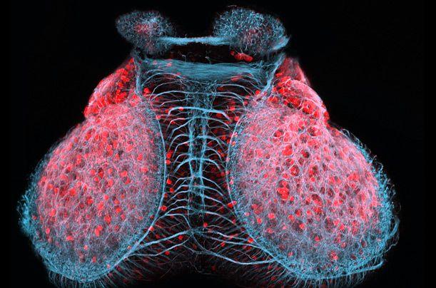
Runner up. Brain zebrafish embryo (zebrafish) three days old. Increased 20 times.
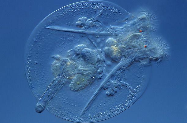
Third place. Rotifer - microscopic marine animal magnified 400 times. From a series of portraits of microorganisms.
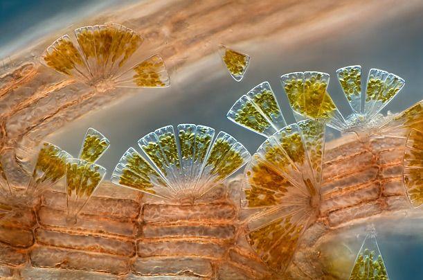
In this photo marine diatoms (a type of phytoplankton) are connected to polysiphonia - red algae. Zoom - 100x.
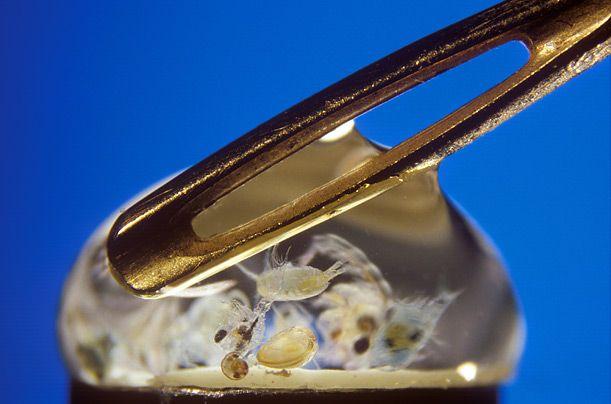
Marine microplankton in seawater. Eye of the needle - to scale.
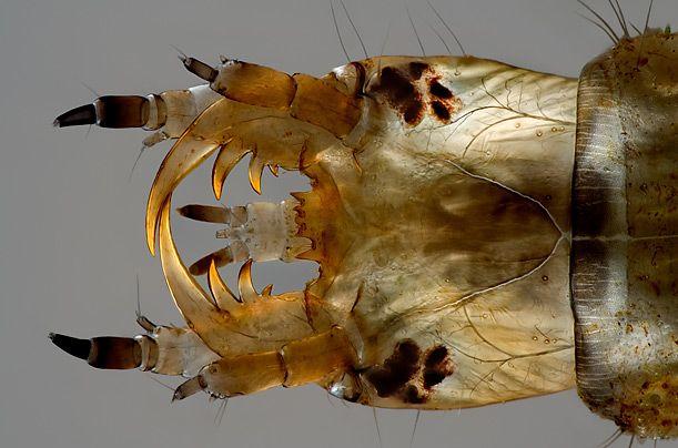
Water scavenger beetle larvae. Complex water beetle head says a lot about his predatory character. Increase - 100x.
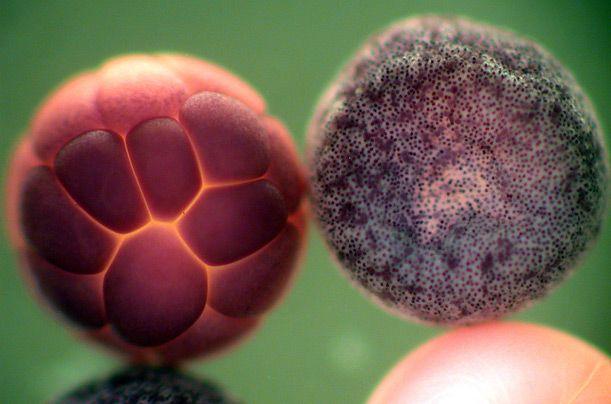
Frog embryos. This image was made to illustrate the distribution of the protein known as Sox3, which plays a crucial role in the early stages of embryonic development of the frog.
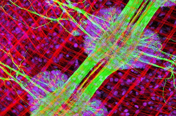
River leech. Abdominal muscles and nerve cord river leeches increased 25 times.
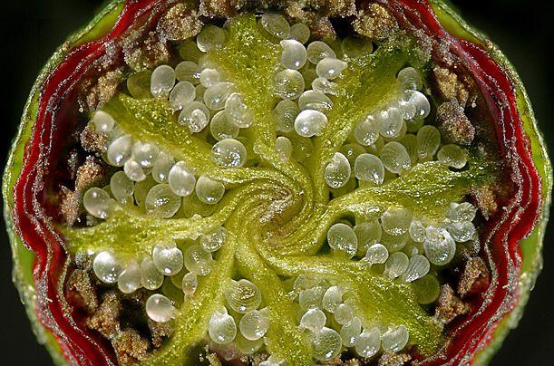
Grain cultivation increased by 20 times.
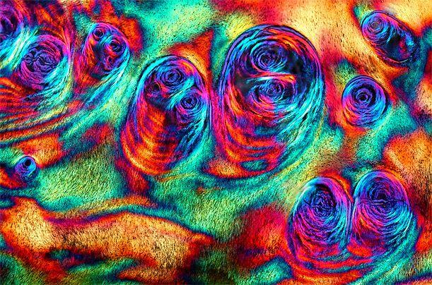
Slice of ivory. In order to show the structure of ivory, the photographer put together with 16 separate images.
via rykun