4114
Scientific, medical illustration and animation: how doctors and scientists to communicate with each other, the students and the rest of humanity
This post we want to give an overview of the area in which we work. In the United States, Europe, Canada and Australia, the public is aware of it, because the area itself is a scientific and medical illustrations developed much better than us. We recall the history of the problem, describe how things are now, what tasks are the main studio of Biomedical Imaging, tell about the organization that this market is examined and supervised, as well as share some statistics.
What's this all about? From drawing to three-dimensional animation of corpses molecules and viruses h4> Our body about 206 bones of different shapes and sizes, over 600 muscles and more difficult to count the number of nerves, blood vessels, and especially all kinds of cells belonging to different tissues and organs. If you delve stronger picture opens a huge amount of protein, RNA and other molecules, each of which performs a specific function, and small disturbances can have a dramatic effect on the life of the organism. Suffice it to recall the prions that are due to incorrect folding of the molecule can cause serious illness.
Living systems are so complex that it is impossible to describe them in detail, using words or formulas. Nevertheless, the description is essential for understanding the mechanisms and causes of the failure of certain biological structures. To solve the problems of descriptive doctors and natural scientists from ancient times learned to make the correct drawings and diagrams as the structure of our own body and the surrounding plants and animals that can benefit us or harm. If in the time of Galen medical and scientific illustration was naive and not exactly that, by the way did not stop his work to be claimed more than 1,300 years, starting with the Andreas Vesalius , she began to take shape as a discipline and an important tool in the training and work of doctors and scientists.
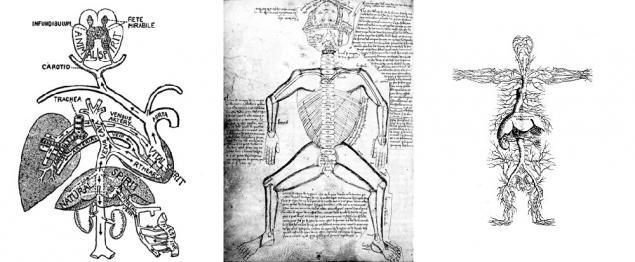
Examples of early medical illustrations. (Источник). I>
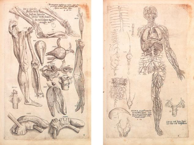
The parts of the human body depicted Andreas Vesalius. Page from the book De Humani Corporis Fabrica, 1543 (источник).
Illustration of how the scientific method has received a new impetus to the development after began to appear first magnifying devices, and undertook business Guk , Leeuwenhoek , Malpighi , Grew and other naturalists, begin to describe the microcosm and prepare the ground for the emergence of the cell theory and understanding microscopic anatomy of living objects.
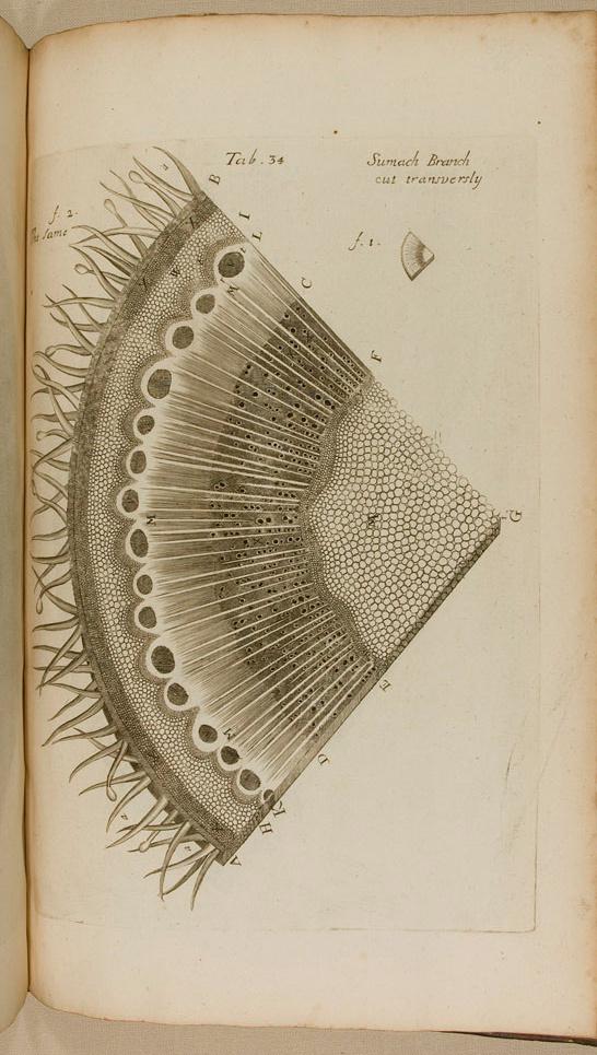
The cut stem of the plant. Author illustration Nehemiah Grew. From the book "The Anatomy of Plants. With an Idea of a Philosophical History of Plants, and Several Other Lectures, Read Before the Royal Society ". 1682. ( Source ) i>
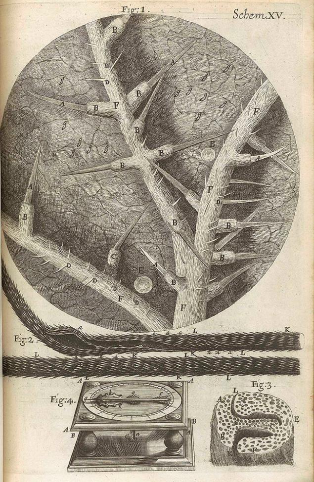
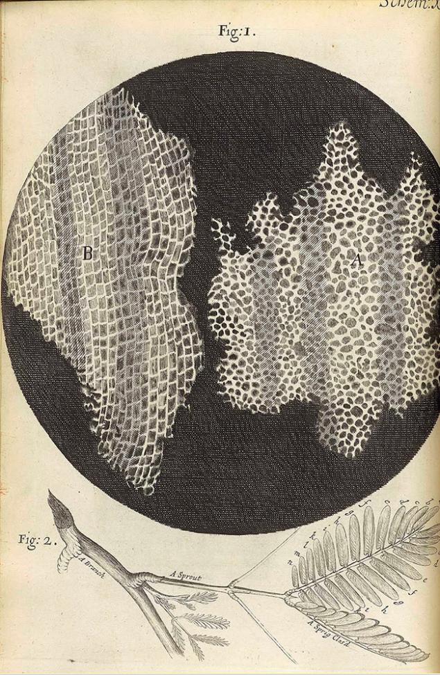
Illustrations Robert Hooke, included in the book Micrographia, published in 1665 ( source ). With these figures living cells get their name and attracted the attention of researchers. I>
Currently biology students in the learning process is still sketched microscopic preparations and dissected with a scalpel cockroaches, but scientific illustration for quite some time became a separate complex discipline that decides not primarily research, and communication and educational tasks. The relevance of this kind of activity is growing, as the volume and complexity of scientific and medical knowledge is growing faster than the ability of people to understand it, and diagrams, models and animations make it possible to study the complex is much more efficient. If a doctor in the XVI century and part medical illustrator at night walking around the cemetery in search of bodies for dissection and sketches, but now such radical approaches are required, and the employment of experts from a wide range of scientists to professionals in graphic design, three-dimensional modeling, computer animation. Of course, the enormous progress in science and the scientific method plays a major role in the evolution of medical illustration. Electron microscopy, X-ray analysis and molecular dynamics have shifted the attention of specialists in this sphere of anatomy and microbiology to molecular medicine, cell biology and biochemistry. Thanks to new techniques become possible to identify and demonstrate structure and mechanism of individual molecules , large молекулярных complexes and even viruses .
The protein kinesin that moves the cell vesicle along microtubules. Studio XVIVO . 2013. I>
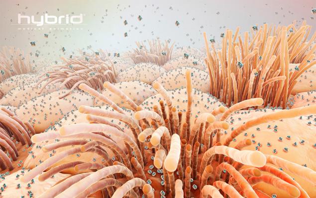
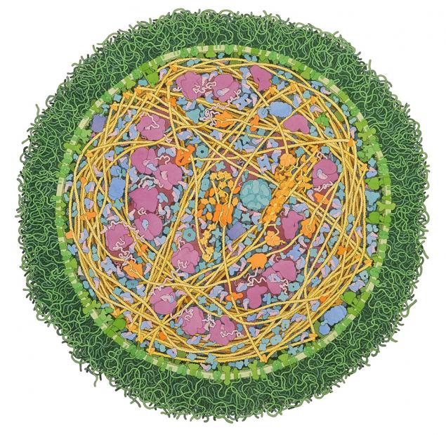
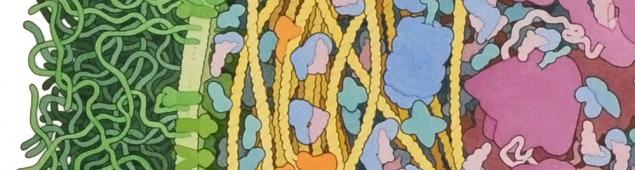
Influenza virus particles on the surface of epithelial cells of the upper respiratory tract. Studio Hybrid .
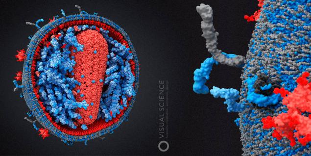
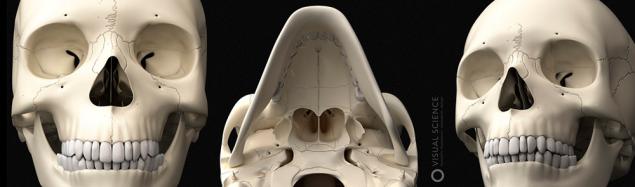
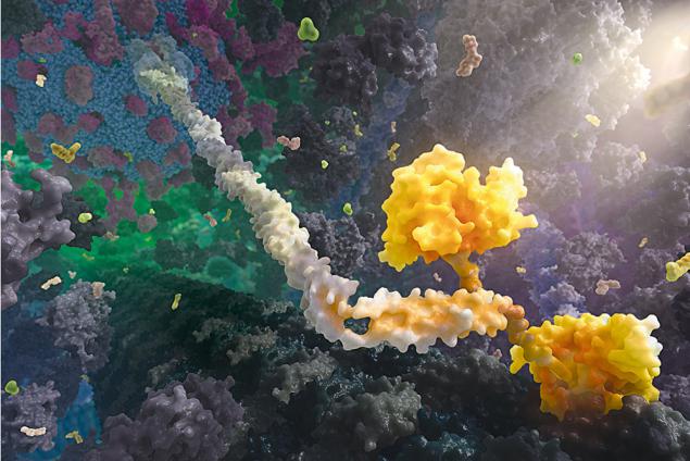
Internal structure human immunodeficiency virus . Studio Visual Science. 2010.


Who needs scientific and medical illustrators? H4> Many high-tech enterprises, whether pharmaceutical companies, companies producing medical equipment, or private clinics need to inform potential customers, partners or investors about what the nature of their development and services offered. Moreover, if they come in a design studio generalist with an order for production of presentation materials, website or animations will likely receive adequate result will be very labor intensive and time consuming. The reason is that the usual designer or illustrator can not have the slightest idea about how, for example, are arranged based nanoparticles copolymer D, L-lactide and glycolide, or affect whether the fracture Le Faure first type ethmoid. Of course, you can embark on a long explanation, scatter references of the designers of atlases and links to Wikipedia, but this approach seriously hinders the process and at high energy-consuming does not guarantee the desired result. At the same time taken by a specialist client who "teaches" the designer, the number of iterations and revisions, make the total cost of this approach is very high. It is therefore formed a specific niche biomedical illustration, animation and design, which is usually either individual experts or teams combine expertise in graphic design, medicine and science.
In addition to high-tech companies in the high technology design, of course, require universities and other educational institutions and projects. According to the latest report of the American Medical Association illustrators , about 39% of the professionals in this field work at a constant rate in various universities and academic institutions. From known examples David Godsell , working in Scripps in California, группу medical animation at the Institute of Medical Research Walter and Eliza Hall in Melbourne, as well as Graham Johnson , develops an algorithm visualization and modeling of molecules and cellular structures of the University of California at San Francisco.
The internal structure of Mycoplasma mycoides. David Godsell. 2011. I>
These illustrations are very impressive in their detail. Notably, all of them are made in pencil and watercolor. David outlines the contours of the molecules in the molecular visualization program and then manually creates a pattern. The disadvantage of this approach is that Godsell as an artist can interpret in their own forms of some objects, without delving into the scientific data that affects the scientific credibility. Plus - that in this way you can do a lot of illustrations of even very large objects in a short time.
Slightly more than one third of medical illustrators as clients and employers have the pharm companies, medical centers and clinics, publishing houses, law firms, specializing in health care (often in litigation, you may want to illustrate, for example, getting a man травму ), as well as advertising and design studio. The rest of the respondents were employed by the Association of Professionals in nonprofit organizations and foundations (18%), and also worked on some government offices (7%).
According to the study, in 2009 the average salary of medical illustrators was $ 61,000 per year, with a range from $ 27,000 to $ 150,000. Studio executives and art directors earned more ($ 93,000 and $ 75,000 per year, respectively). Graphic designers, animators and developers of multimedia content to earn about $ 47,000 to $ 58,000.
Total employment was analyzed more than 550 professional members of the association.
Part of Biomedical Imaging is a scientific illustration and animation. Specificity is the most accurate work with the original data - often the results of experiments or simulations in silico. The tasks are the same - communication in a professional environment (starting with a certain level of well-known authors), presentation, illustrate scientific publications, textbooks and other educational materials.
Scientists in everyday tasks and self nice find a common language with each other. Presentations at conferences usually are primitive in terms of graphic design and graphics, but in most cases they surely solve your problem, since the audience is prepared and relatively undemanding to the level of supply. Difficulties arise when scientists have to explain something to the audience with a different level of immersion in the subject, such as students, or even from other areas, such as business, in the case of investors or partners at the stage of commercialization and popularization of development. In this situation, it's much more difficult to explain, and qualitative visual materials are needed, in order not to be limited to a set of arguments Richard Dawkins in this passage: coub.com / view / 1j2dqvn .


Biomedical illustration now h4> The market biomedical illustration and animation are a few large companies and a lot of smaller players and individual illustrators and designers. Basically specialists of this sphere are concentrated in the US, Canada, and quite a bit in Europe and Australia. One of the leading animation studios, XVivo, became widely known through a large-scale three-dimensional roller Inner Life of the Cell, which demonstrates the molecular processes occurring in the leukocyte when activated during inflammation. The spot was made for the educational program at Harvard in molecular and cell biology, but quickly gained a much wider audience. With its creation, the company has used the work of students at Harvard (rumor - for free).
Many well-known studio did not pay more attention to intracellular processes, and the anatomy and physiology of the human body. Examples of this work demonstrate the Hybrid, Zygote , Nucleus a >, Brian Cristie , Invivo , Anatomyblue , AXS Studio . In this case is not limited animation studios create three-dimensional models of learning, illustrations and applications. This theme can be important not only for education but also for hospitals and clinics .
To get an idea of the field of scientific visualization, you can see the works of the participants of competitions that are held in this area. The most famous of these was the Science and Engineering Visualization Challenge , which is held by the journal Science in conjunction with the National Science Foundation, which we also participated in 2010 and 2011. It focuses on the natural sciences, and the participants are sent as illustrations and infographics and animations, and even educational games. Computer graphics and animation created in the studios of the scientific design also takes presented at competitions SIGGRAPH , whose range of participants is not limited to medical illustration. Effective communication and marketing in the field of medicine and health care assesses the jury, conducted by the Medical Marketing & Media . Another notable, but the dealer several positions contest called Vesalius, and there is a focus on the educational component.
Association of Medical Illustrators, which has already been mentioned, certifies professionals coordinates a kind of trade union initiatives, conducting market research and analytics, and produces, among other things, exists since 1946. They also carry own competition and exhibition at the annual meeting of members, with the works of the winners of which are available on the website of the organization. In the competition, unlike SEVC, focuses on the medical component.
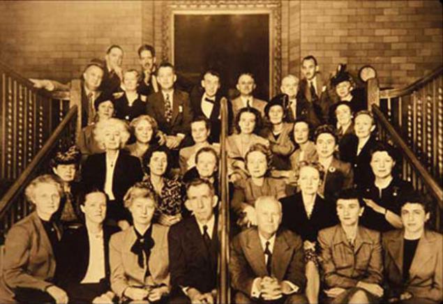
Photos from the first meeting of the AMI. Philadelphia, PA. 1946. (Источник)
Besides the competitions, a slice of the area can give catalog of medical illustrators , which is published annually, or фотобанки, specializing in this subject, which presents hundreds of thousands of pictures, photos from experimental to Sainz art.
If the topic is interesting, in the next posts we will cover a much wider field of communication in biomedical areas of the so-called healthcare communication, part of which is a biomedical illustration and animation. The topic of the next post:
Browse destinations healthcare communication Analysis and recommendations for scientific posters and illustrations of articles readers and authors Habra. Works can be sent to the email mailbox @. Make a post as typed 10-15 The story of the process of creating 3D models of human viruses in Visual Science - Part 2, molecular modeling and 3D < Medical anatomical illustration - history of the study of the human body in the work of illustrators 5 centuries The so-called Sainz art or scientific art table > Only registered users can vote in polls. Sign , please. 82 people have voted. 19 people abstained. 
Source: habrahabr.ru/company/visual-science/blog/225277/


A drop of water and superhydrophobic surface
Intel unveiled a prototype laptop with wireless charging, WiGig and passive cooling