868
Micrographs from the competition Nikon (14 photos)
We bring you the pictures from the photo contest «The Nikon Small World Competition». He will become the premier event for showcasing the beauty and ease of microcosm, the competition celebrates the best "micro-photography", creating beautiful photos at the same time revealing the scientific point of view of the object.
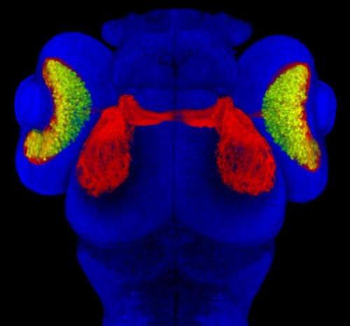
1. John Gaines
University of Utah
Salt Lake City, Utah
3 days after fertilization zebrafish embryo
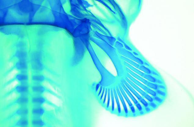
2. Dr. Andrew Gillis
Cambridge University
Cambridge, UK
Pectoral fin embryo Whitespotted bamboo shark
Picture taken through a stereoscopic microscope with fiber-optic lighting
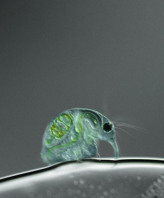
3. The role of Joan
Institute of Biochemistry and Biology
Potsdam, Germany
Daphnia (x100)
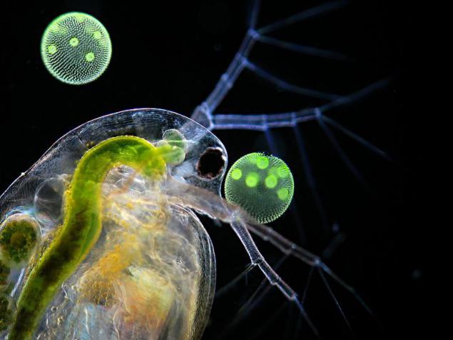
4. Dr. Ralf Wagner
Dusseldorf, Germany
Water flea and round balls of green algae
On a dark background with flash
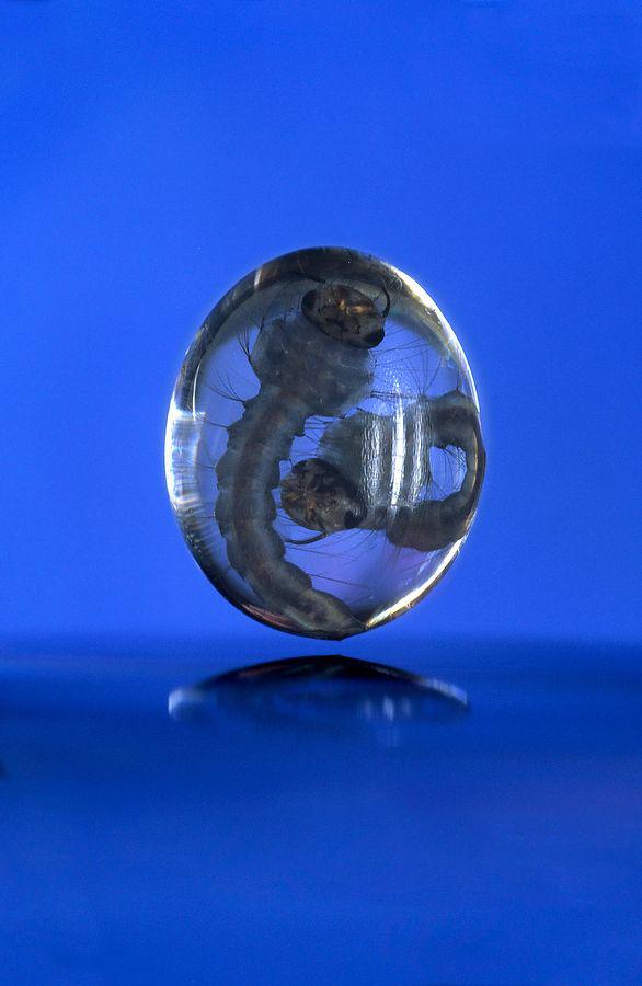
5. Dr. John H. Brackenbury
Cambridge University
Cambridge, UK
Drop of water with a pair of mosquito larvae
Closeup with high-speed laser initiation
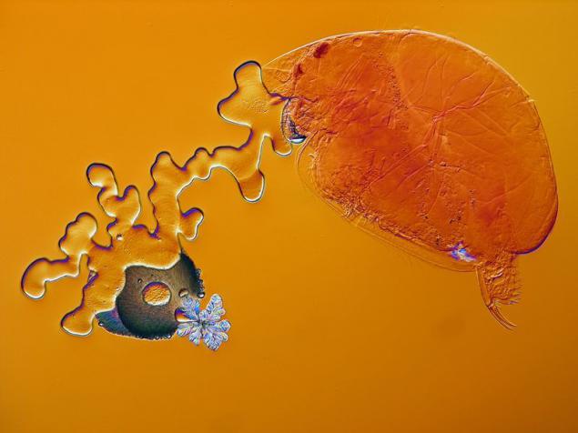
6. Dr. Carlos Alberto Munoz
University of Puerto Rico campus Mayagez
Crustaceans in fir balsam with crystals and other particles
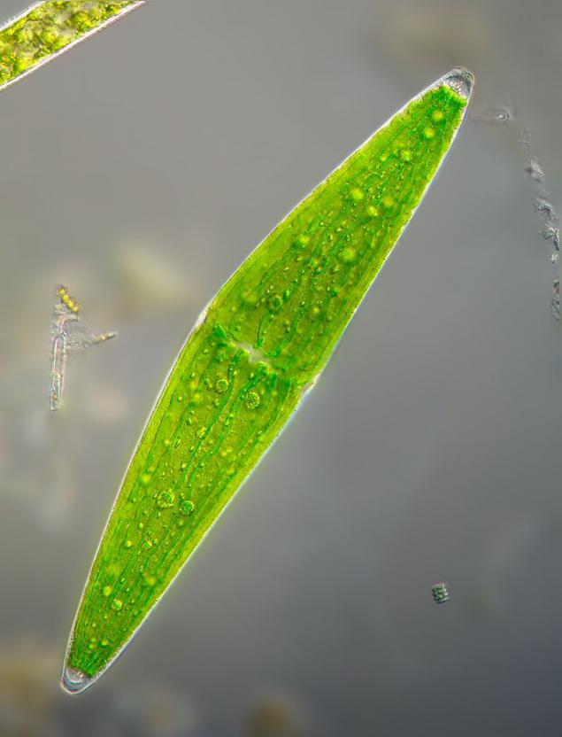
7. Wolfgang Bettighofer
Kiel, Germany
Green algae from the pond marsh
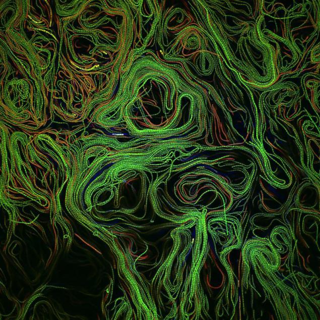
8. Jonathan Franks
University of Pittsburgh
Pittsburgh, PA
Biofilm algae
Autofluorescence, with a common focus
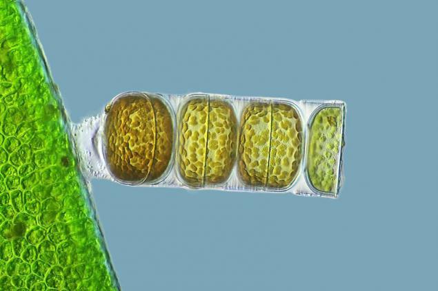
9. Frank Fox
University of Trier
Trier, Rhineland-Palatinate, Germany
Moniliformny red zoster (h320)
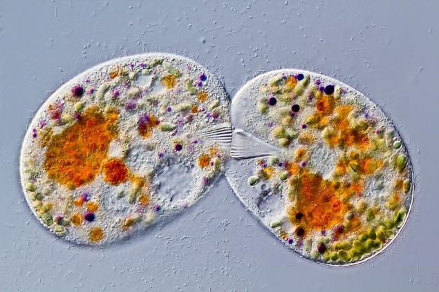
10. Gerd A. Guenter
Dusseldorf, Germany
Freshwater ciliates, conjugation, living species (h630)
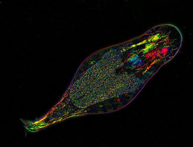
11. Michael Shribak / Dr. Irina Arkhipova
Laboratory Marine Laboratory
Woods Hole, Massachusetts
Rotifer Filodina
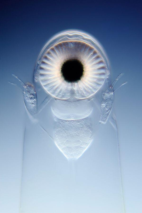
12. Wim baths Egmond
Museum Mikropolitan
Rotterdam, Netherlands
Eyes large water flea
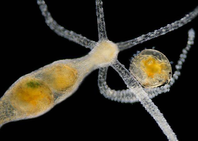
13. Charles Krebs
Charles Krebs Photo
Issaquah, WA, USA
Hydra is devouring water flea
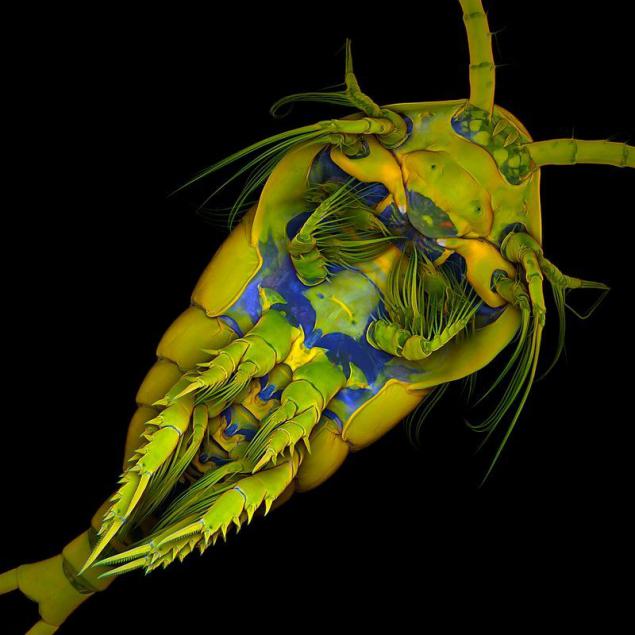
14. Dr. Jean Mishels
Christian-Albrechts University in Kiel
Kiel, Germany
Marine copepod cancer, a bottom view (x10)
1. John Gaines
University of Utah
Salt Lake City, Utah
3 days after fertilization zebrafish embryo

2. Dr. Andrew Gillis
Cambridge University
Cambridge, UK
Pectoral fin embryo Whitespotted bamboo shark
Picture taken through a stereoscopic microscope with fiber-optic lighting

3. The role of Joan
Institute of Biochemistry and Biology
Potsdam, Germany
Daphnia (x100)

4. Dr. Ralf Wagner
Dusseldorf, Germany
Water flea and round balls of green algae
On a dark background with flash

5. Dr. John H. Brackenbury
Cambridge University
Cambridge, UK
Drop of water with a pair of mosquito larvae
Closeup with high-speed laser initiation

6. Dr. Carlos Alberto Munoz
University of Puerto Rico campus Mayagez
Crustaceans in fir balsam with crystals and other particles

7. Wolfgang Bettighofer
Kiel, Germany
Green algae from the pond marsh

8. Jonathan Franks
University of Pittsburgh
Pittsburgh, PA
Biofilm algae
Autofluorescence, with a common focus

9. Frank Fox
University of Trier
Trier, Rhineland-Palatinate, Germany
Moniliformny red zoster (h320)

10. Gerd A. Guenter
Dusseldorf, Germany
Freshwater ciliates, conjugation, living species (h630)

11. Michael Shribak / Dr. Irina Arkhipova
Laboratory Marine Laboratory
Woods Hole, Massachusetts
Rotifer Filodina

12. Wim baths Egmond
Museum Mikropolitan
Rotterdam, Netherlands
Eyes large water flea

13. Charles Krebs
Charles Krebs Photo
Issaquah, WA, USA
Hydra is devouring water flea

14. Dr. Jean Mishels
Christian-Albrechts University in Kiel
Kiel, Germany
Marine copepod cancer, a bottom view (x10)

