2798
Part 2. How many Mbit / s can be passed through the optic nerve and the retina at a resolution? Some theory

Previous publication , dedicated to the technologies of laser vision correction has been met with great interest that I honestly did not expect. That's why I decided to continue the story in the form of a cycle in which we will look at more technology underlying the laser eye surgery. If you expected to see themselves directly lasers in this article - I have to disappoint you a bit. I've been trying to get around a biological theme, but eventually realized that I could not talk about laser vision correction, without revealing the foundations of the structure and functioning of our vision.
I will try to consider the human eye through the lens of IT. If someone is not very interesting to read the part dedicated to the biological aspects of - do not worry. Just skip sections, starting with the optical system of the eye, and immediately proceed to the traditional competition from our girls. However, I still recommend you read this material to understand better the next article, we will consider LASIK, Femto-LASIK, ReLEx SMILE and other methods of laser eye surgery.
Is the mood to find out what exactly do these strange people in white coats, thoughtfully looking at the results of your survey? Do you want to learn a little about the new unique natural gift - view? Then welcome under habracut. As usual - many illustrations and traffic (≈5 MB).
Contents:
Vision and IT
small entry retinal megabits compare with the camera? The optical system of the eye
cornea front and rear cameras iris and pupil lens Vitreous Why do we see the bad? Refractive disorders
Myopia spasm of accommodation < a href = "# ancAstigmat"> Astigmatism Hyperopia Presbyopia Bonus
Mini interview with laser surgery next competition from our girls
Instead of joining
Eye - sophisticated optical instrument created by nature. The man was in many ways just an imitator, producing their creations. Optics has come a long way as the science of Newton's experiments with a prism to developing unique lasers, night vision devices, and other interesting things. However, a closer look at what is largely inspired mankind to study and became a prototype for many modern things. The human eye.
retina and optic nerve
Any device capable of detecting light usually has one or another version of the image sensor. Its biological analogue is the retina of the eye.
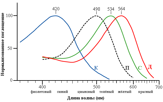
Normalized graphic sensitivity of human cells different types of cones (A, C, D) and rod cells (R) to different parts of the spectrum. The axis of the wavelength on this logarithmic graph. I>
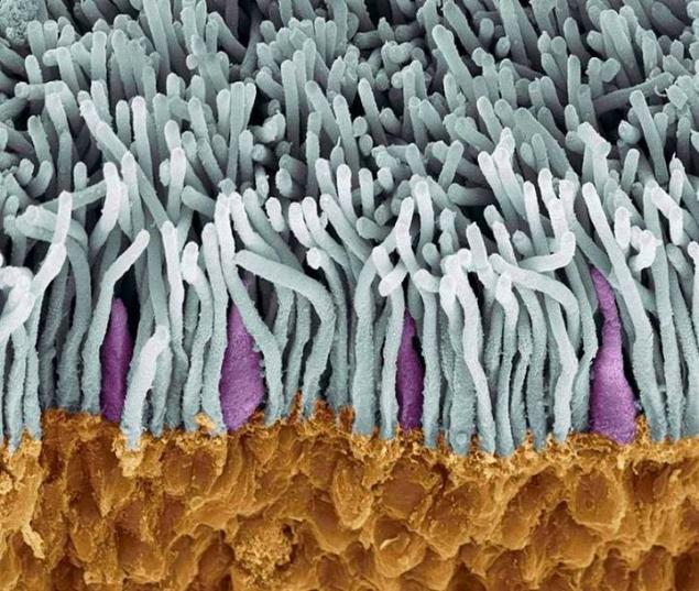
An electron micrograph of the retina. Sticks are gray and purple cones. In the illustration, you can see a clear predominance of rods over the cones monochromatic i>
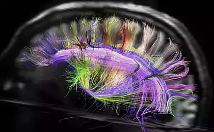
For example, the retina of the human eye has about 7-8 million cones are responsible for color vision, and about 120 million rods (black-and-white vision). Not quite be correct to equate the flask / stick to the pixel, but, according to rough estimates, their number corresponds approximately 250 megapixels for panoramas with both eyes. After excitation of light-sensitive cells of the retina, the signal to be transmitted to our biological CPU / GPU. This function successfully executes the optic nerve. The bottleneck in the maximum frequency is limited by "personnel", transferred the body of view, is - latency nerve synapses i> (land connections between neurons, where the pulse is transmitted by emission and capture of chemicals). According to various estimates, is about 100-150 Hz, which is the limit of the speed of image transmission in the visual cortex of the brain. The number of nerve fibers in the optic nerve is approximately 1 200 000. If we assume that one fiber per cycle may transmit one bit of information, the combined capacity of the optic nerve is approximately equal to 1.2 x 10 6 sup> * 150 = 180 bits Mbit / s. Solid stream. Our brain has to handle the total flow of 360 Mbit / s only on the visual analyzer. But there are also hearing, smell, touch, temperature and pain receptors, sense of balance, a feeling of proprioceptive (feeling your own body in space). All of this load, not including heaps of other features, fit only 25 Watt TDP.
Here, incidentally, covered a number of issues that lie in the field of bioinformatics. For example, it is not clear how ≈125 * 10 6 sup> cells receptor can transmit information in ≈1.2 * 10 6 sup> conducting neurons. That is, before the transfer takes place in the brain is a kind of filtering and pre-processing of visual information. By the way, it follows that the "beat" the retina is unable to deliver a 1.2 megapixel camera. Another thing is that as a result of processing a series of "snapshots" in the brain formed a much more clear picture of what is happening. Another interesting feature of the human eye is the fact that we always see past i>. The delay in carrying out a nerve impulse to the center is variously estimated about 150-180 ms. That is why, by the way, in professional sports is considered premature to launch a breakthrough athlete in the interval from 0 to 100 ms. It is believed that a person could not due to the physiological limitations have time to react so quickly. It should be noted that these values can vary within certain limits depending on the stress, psycho-emotional state, the level of neurotransmitters, but the overall picture is stable enough.
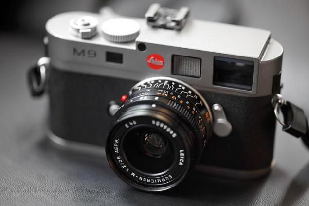
compare with the camera?
Sensitivity may undergo substantial widest range - from normal sight with illumination of 25,000 lux (bright noon) prior to the registration of individual photons in the pitch dark at maximum adaptation. The dynamic range of the eye and strikes against the backdrop of traditional cameras - about 24 f-stop! For comparison, the maximum dynamic range is among the photographs of black and white film - about 10 f-stops. Color film has a range of about 7, and the average matrix of the modern camera 4 to 6. Thus, the dynamic range of the eye more than the average camera in 2 (24-6) sup> = 262 144 times b >.
It is worth noting that this adaptation takes time. The rate of addiction over the transition to a bright room - only a few seconds. In the transition to the dark time is extended to several minutes. This is due primarily to the need ocherel synthesis destroyed rhodopsin i>, the visual protein that is directly responsible for the emergence of the visual field. Luminous flux is regulated as well as in a camera - Aperture. This function is performed by the iris, and the hole in which the pupil is called. Knowing that the focal length of the eye is about 22 mm, you can count F-number abbr> i> for the pupil: to maximize pupil 8mm - 22 / 8 = 2.75, for the most narrow - 22 / 1.1 = 20.
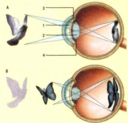
In fact, our eyes - wide-angle lens with its inherent distortions in the periphery. However, the reconstruction of three-dimensional images in the brain is compensated. Also, we do not notice Blind Spot abbr>, even though it is quite noticeable angular dimensions (about 1.3º).
optical system of the eye
For proper focusing of the rays we are not only the size and shape of the elements, but their refractive index. You have to understand that each light guide element has its own, well-defined factors. Incidentally, this explains why we, unlike fish, because of poor visibility under water - our eyes have evolved as a body to provide a clear view on the land. All this is because the power of the lens depends on the refractive index.

Glass lens loses much of its ability to focus light on Wednesday when transferring to another IOR abbr> i>
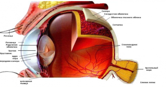
The eye is a finely balanced optical system. The slightest change in the ratio of its members is likely to disrupt the final image on the retina. Imagine for a moment that you have taken and put in manual mode arbitrary focus on the camera lens. You agree that a good and clear picture in this way you will not get. Let us consider in detail which way overcomes the light towards the retina.
The optical system of the eye. Indicate the main dimensions and the refractive indices of the media
cornea
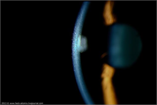
corneal slit ophthalmoscopy i>
The cornea with a large increase
The first element of meeting the onslaught is not too friendly environment - the cornea. The cornea is the eye to the outer shell and a protective and light-refracting function. It is this coating of the eye is facing a permanent negative impact of the dust specks, sunlight and other factors. The corneal epithelium is constantly updated to ensure its integrity and barrier properties. Interestingly, the cornea has no blood vessels in their optical part - it would violate transparency. Power and gas exchange of these areas is carried out by diffusion due to the tear fluid outside and the aqueous humor of the anterior chamber inside.
front and rear cameras
The picture is visible anterior chamber, located just below the cornea and filled with a special liquid - the aqueous humor. The rear camera is located behind the iris and the pupil.
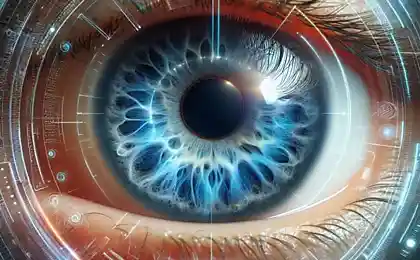
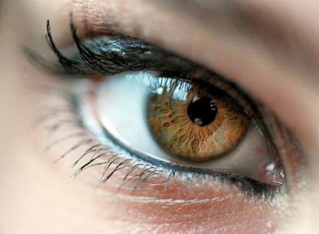
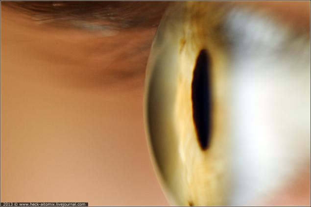
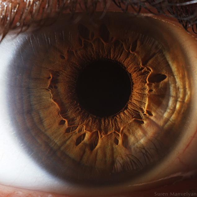
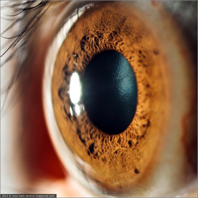
iris and pupil
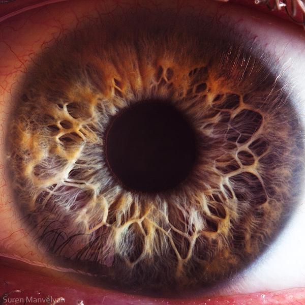
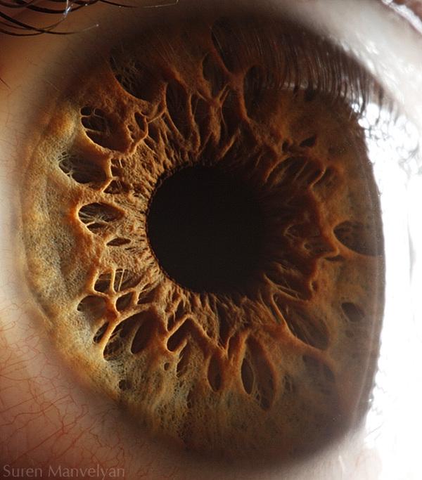
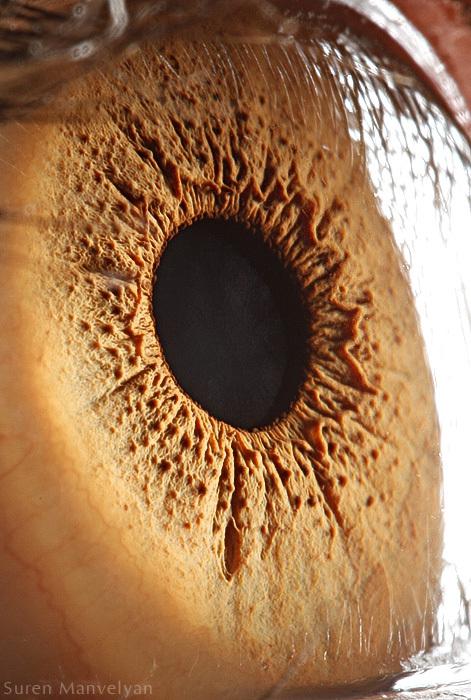
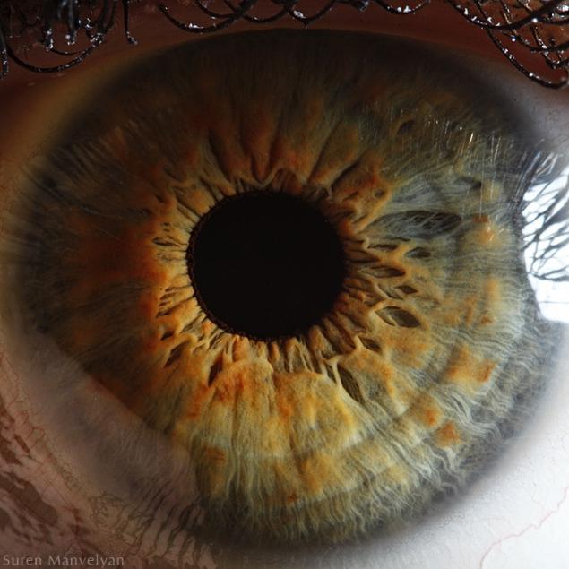
Each person - the owner of a unique in its beauty, and the palette of the iris pattern. For IT-specialists Iris is primarily an important element for biometric identification. For the doctor - this is a very important element, which is similar to the diaphragm controls the flow of incoming light. A hole in the iris and is called pupil i>. Few realize that the iris has a fairly pronounced three-dimensional pattern, which can show the photographs to put right light. Often used polarizing filters to suppress reflections from the cornea.
In fact, many have never thought that the relationship глубины sharpness and diameter of the aperture extends to the pupil. In the dark depth of field decreases sharply, as it increases the iris-pupil.
More beautiful pictures of the iris.
lens
The lens is the unique biological lens, which has a number of very important properties. One of the most important - the ability to change its curvature under the action of the ciliary muscle. This process is known as accommodation and allows to focus on distant or at very close objects. Accommodative possibilities of optical system of the eye of a young man is ~ 14 diopters, gradually decrease with age and 60-65 years of virtually lost. By the way, thanks to the lens optical system of the eye is so compact in comparison with, say, SLR cameras.
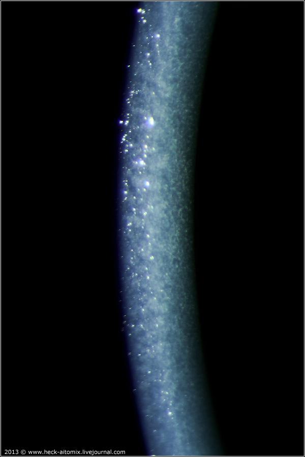
The reflection of the front of the lens at the slit microscopy
Vitreous
Contrary to popular belief, that the eye is filled with fluid that can flow at the slightest puncture, the bulk of the eye takes the vitreous. This substance is more like a viscous gel, whose mechanical properties are determined primarily hyaluronic acid. The main function of the vitreous - the maintenance of stable form the eyes, giving it the necessary elasticity. Also, the vitreous body holds itself through and refracts light.
Refractive disorders
All these elements belong to the refractive system of the eye. That is why any violations associated with them are called refractive i>. This type of pathology is interesting because we are able to restore the correct path of the rays, acting on the wrong item, which was the cause of the disease. For example, the use of points as an additional lens corrects nearsightedness, the cause of which was the increase in the central optical axis of the eye. Consider next the main problems associated with the light refraction.
Habrazhitel ansaril3 suggested adding an article in a physical justification of such violations. Unfortunately, my medical degree does not allow me to fully understand the meaning of these things, but I'll leave the link for those who are interested.
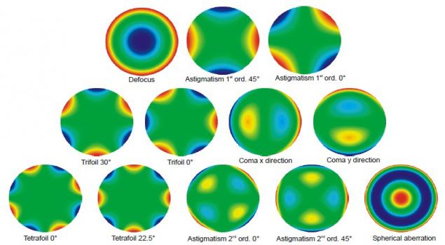
Zernike polynomials and wave aberration :
Nearsightedness (myopia)
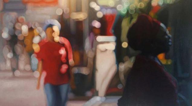
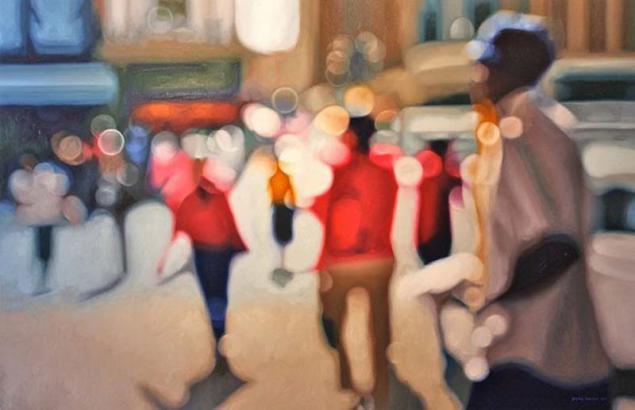
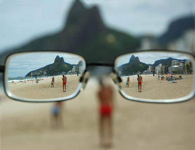
Beach many experts in the field of IT. Before we talk about the causes of this disease, I want to refer briefly to the art. Philip Barlow - a talented South African artist who was able to reflect in his work through the eyes of a short-sighted person.
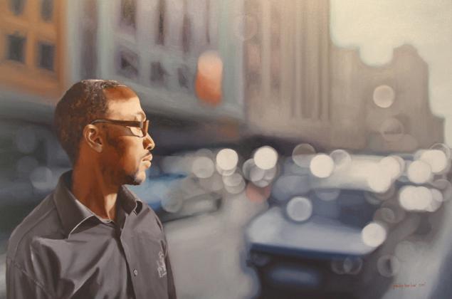
A little more works by this author


The cause of myopia is increasing the size of the eyeball along its optical axis:
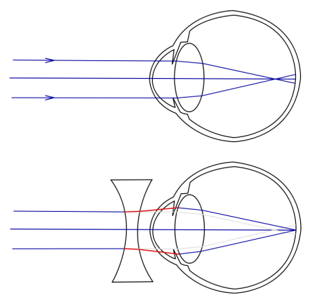
This disease is often most pronounced in adolescence, a period of sharp growth of the organism. There are observations that linked excessive stretching of the eye with genetic defects in collagen synthesis. Collagen - the structural protein, which is important in the formation of connective tissue. When it comes to excessive elasticity and disproportionate growth of the eyeball. In the growth stage, to stop the further growth of short-sightedness may use sclerosis and kollagenoplastiku i>. The essence of these methods is increasing at the strength of the outer membrane of the eye - sclera i> - by implanting a special material. Treatment of this disease is non-operational methods (glasses, contact lenses) and operational (different types of laser vision correction).
We have the opportunity to correct this problem Technology Femto-LASIK, in the coming weeks we will also be able to work on the technology ReLEx SMILE. We use the latest generation of ophthalmic lasers:
For Femto-LASIK is Amaris 750S is powered by Schwind (excimer laser for the correction itself) and VisuMax from Zeiss (femto-laser for cutting out the flap)
For ReLEx SMILE is only VisuMax (technology means only perform all the manipulations on it in greater detail in the next article)
spasm of accommodation
It is necessary to distinguish between the true myopia, and the so-called cyclospasm i>, also known as false myopia. I think that in view of the profession, many of us spend a lot of time looking at the monitor continuously. As we said earlier, is responsible for the accommodation of the ciliary muscle, which deforms the lens as needed. At constant eyestrain when the focus of a long period of time is near, this muscle spasms and tests can not relax. As a result, the eye loses its ability to properly focus on objects in the distance, but this is due to a temporary phenomenon in terms of accommodation and not to change the form of the eye. In this situation, appoint special drugs that cause temporary paralysis of the ciliary muscle, helping it to relax (tropicamide, atropine, and others). It is worth the visual gymnastics and hygiene of labor (workplace lighting, breaks, etc.)
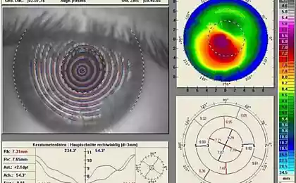
Astigmatism
This pathology is often combined with others. Its cause is asymmetrical curvature of the cornea or lens. The result is a different refraction with respect to different axes. As a result, a person can clearly see, for example, horizontal lines and the vertical will be blurred.
A little bit more about astigmatism

Type of test миры the eyes of a person suffering from astigmatism i>

The test of the world in its original form.
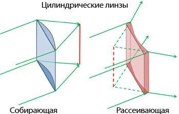
Treatment - the use of special cylindrical lenses in the glasses or laser vision correction, during which will be adjusted, and this pathology.
farsightedness (hyperopia)
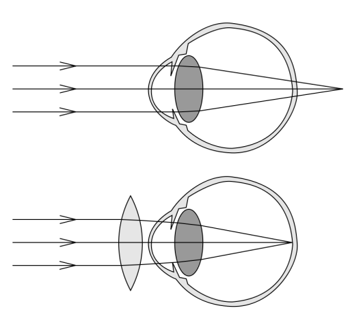
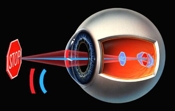
Status opposite of myopia. The optical axis of the eye is shorter than they should be, with the result that the image is focused behind the retina.
This disease is often confused with the presbyopia i> (age farsightedness)
Treatment of ophthalmic lasers substantially similar to the treatment of myopia.
Presbyopia (age farsightedness)
The peculiarity of the refractive disorders that a person loses the ability to age for accommodation.