479
How to interest the student of natural Sciences
Modern science education in the majority of schools serve one purpose: to discourage student interest in the subject. With almost a decade of experience creating graphics for the leading publishers of scientific literature, illustrations, published in The New York Times and The Washington Post and even images for the presentations of Nobel laureates, we are determined to combat this problem.
In this post outlined a story about how we helped the associates from the laboratories of the Polytechnic Museum to develop the usual school format educational posters, providing students the app with augmented reality. In the process we got a detailed three-dimensional model of the prepared frog, based on data from computer tomography, to visualize a gene and its corresponding protein in one scale, and modeled interactive atom, allowing to see the orbitals of different shapes and sizes.
Forty eight million five hundred eighty nine thousand three hundred eighty nine
Posters and posters is a common thing for education, has long been integrated into the educational process. Despite the abundance of digital boards and other innovative equipment, posters are still used in schools but often in need of serious updating.
Obsolescence is a good excuse to the teacher-the enthusiast replaced the ancient poster on the more relevant (besides containing a surprise). We have no illusions that only one poster can solve the problem of motivation and engagement of pupils, but a pilot project in which the poster is a “window” to access the advanced content in a familiar student environment tablet or smartphone, seems to us interesting to evaluate the potential of such a venture.
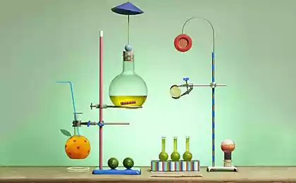
Flat illustration may be informative and attractive, but for a two-dimensional image is not easy to really in detail and to demonstrate the device complex objects, which even in the school curriculum quite a lot (from the structure of molecules or of living creatures to pilot plant or atomic orbitals).
On a complex object I want to see from different sides to remove layer by layer, or to see the individual parts. This is possible when you combine an educational poster and mobile application created using augmented reality technology, which we did. In addition, the app contains “trimmed” the electronic version of the posters that adapt the content of the poster to the mobile device.
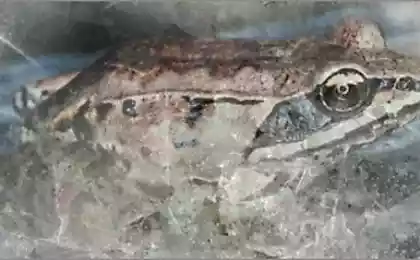
The anatomy of a frog On the one hand, this theme has been in production for decades: there are lots of books, illustrations and videos demonstrating how the frog; available diagrams of individual organ systems and of course posters.
Nineteen million four hundred eighty six thousand one hundred seventy five
Existing illustrations are primarily done in the style of classical monochrome drawing. This allows you to thematisiert material, which is often good, but in most cases these are illustrations contain many inaccuracies and incorrectly reflect reality.
Surprisingly, the most complete Russian-language guide to the anatomy of the common frogs (the book by P. V. Terent'eva “Frog” from the “Laboratory animals”) is illustrated very poorly, and in fact contains many not the most obvious points, which to understand the topic without zoologists working in vertebrates, almost no chance. When it comes to the mutual position of movable parts of the body or, for example, blood vessels relative to the skeleton, you should dissect an animal to understand the issue.
Sixty four million one hundred seventy five thousand fifteen
The video preparation has its own problems: they can show is not all that is required (the most difficult in this plan are submitted to the circulatory and nervous system).
The second key problem of the existing school manuals is that they are unable to attract and retain the attention of the student, experienced works the best specialists games, advertising and film industry, the controlling disparate budgets while rendering even the simplest of objects and phenomena.
Our task in this case was the creation of the most accurate and close to reality the benefits that would be useful for familiarization with the structure of all organ systems of a frog. And, of course, we wanted to “recapture the smell of mothballs” from biology as a discipline – at least in this particular case. Traditionally, we use hybrid approaches, which are based on three-dimensional graphics. No illusions, and plans for what we will be able to draw and simulate a frog on existing atlases and illustrations we have not had this approach obviously looked like a dead end. We have therefore set ourselves the task to obtain the most accurate tomographic scans of the frog.
Search the frog was not easy. First, it turned out that by the end of may, when we needed the animals, they have already finished breeding, and to find them in suburban forests themselves have failed. Besides, zoologists and physiologists, who annually teach students on frogs, say a sharp decline in their numbers in the forests around Moscow, which the fishers of frogs harvested animals away from the city. Search for pet shops and bird markets also returned no results – some for the same seasonal reasons.
I had to look for people who are directly involved in catching frogs for terrariums, shops and educational institutions. In the end we got the desired three members of the species Rana temporaria in the chilled and sleepy.
Ninety one million seven hundred eighty five thousand two hundred forty one
Modern methods of imaging allow to scan with a resolution up to 20 microns (micron is one ten thousandth of a centimeter). This means that approximately vosmiseriynogo frog will have approximately 4,000 horizontal “slices”.
In our case the layer was somewhat smaller, but still their number was more than sufficient to build a truly accurate model, where one can distinguish, for example, a finger phalanx of the animal and even quite small vessels. Only this approach ensures the accuracy of relative position of all organs and organ systems in the final model. The appropriate resolution of the scanners used to estimate defects – for example, in electronics.
Thirty eight million nine hundred sixty five thousand eight hundred forty six
One of these x-ray scanners we were able to access. It would seem that after this, everything should be pretty straightforward: put the animal into the device, get the scan. In fact there is a problem of contrast of internal organs: in other words, a “head” MRI allows us to distinguish only very dense tissue, like bone, and soft tissues on the images merge.
This problem is widespread in connection with what researchers have already tested various techniques of treatment, based on the difference in the ability of different tissues to pass and accumulate contrast agents (usually in this role are substances containing atoms of relatively heavy elements – Halogens heavy metals). We had to thoroughly study the methodical scientific literature, and then to address to colleagues from the Department of chemistry for assistance in the preparation of the necessary reagents. But in this case, some tasks remained unresolved.
In particular, bones, muscle tissue, cavities and organs after treatment become sufficient contrast, and circulatory system no. As a result of work we had to adapt existing protocols and to develop a new technique that solves this problem. At this stage, our team was joined by zoologists from the Biological faculty of MSU, who helped to dissect the animal and to carry out the alternative injection of contrast agent into the circulatory system prior to imaging.
Fifty four million nine hundred forty two thousand eight hundred eighteen
As a result, a combination of software for the analysis of computer tomograms with popular programs for three-dimensional modeling, we were able to obtain a complete model of a frog together with the models of all its main internal organs, blood vessels and skeleton. It took about a month of hard work on the markup, “cleaning” and additional 3D modeling.
Sixty one million thirty three thousand three hundred eighty one
Thirty three million five hundred forty one thousand one hundred ninety one
Twenty three million one hundred seventy three thousand six hundred thirty seven
The image on the poster was created based on the obtained model, and a special app with augmented reality for modern devices based on iOS and Android it is possible to view the model from different sides to see the structures that are not visible on static illustrations.
Such a comprehensive approach to school poster may seem redundant.
But, first, the example three-dimensional models of viruses that were not created for schools, we have noticed that students appreciate when they are treated in the same way as with older people. They love it when they are offered together to understand something at first glance complex, inviting to rise to the level of the scientist, instead, to consider the scheme of unknown artists with the template definitions from the textbook.
Second, we are under no illusions that all books should be illustrated this way, but I believe that in school there is a place for bright and interesting “entry points” in the theme where we can exert maximum effort and still captivate student biology, chemistry or physics.
Third, anticipating the issues of green and animal welfare activists, we want to convey the idea that such a detailed 3D model can be the basis for educational courses on the anatomy of a new generation, in which General studies students not planning to continue to work with experimental animals, don't have pointless to destroy hundreds and hundreds of frogs, rats and pigeons just to be considered their insides. published
P. S. And remember, just changing your mind - together we change the world! ©
Join us in Facebook , Vkontakte, Odnoklassniki
Source: geektimes.ru/company/visual-science/blog/266866/
In this post outlined a story about how we helped the associates from the laboratories of the Polytechnic Museum to develop the usual school format educational posters, providing students the app with augmented reality. In the process we got a detailed three-dimensional model of the prepared frog, based on data from computer tomography, to visualize a gene and its corresponding protein in one scale, and modeled interactive atom, allowing to see the orbitals of different shapes and sizes.
Forty eight million five hundred eighty nine thousand three hundred eighty nine
Posters and posters is a common thing for education, has long been integrated into the educational process. Despite the abundance of digital boards and other innovative equipment, posters are still used in schools but often in need of serious updating.
Obsolescence is a good excuse to the teacher-the enthusiast replaced the ancient poster on the more relevant (besides containing a surprise). We have no illusions that only one poster can solve the problem of motivation and engagement of pupils, but a pilot project in which the poster is a “window” to access the advanced content in a familiar student environment tablet or smartphone, seems to us interesting to evaluate the potential of such a venture.
Flat illustration may be informative and attractive, but for a two-dimensional image is not easy to really in detail and to demonstrate the device complex objects, which even in the school curriculum quite a lot (from the structure of molecules or of living creatures to pilot plant or atomic orbitals).
On a complex object I want to see from different sides to remove layer by layer, or to see the individual parts. This is possible when you combine an educational poster and mobile application created using augmented reality technology, which we did. In addition, the app contains “trimmed” the electronic version of the posters that adapt the content of the poster to the mobile device.
The anatomy of a frog On the one hand, this theme has been in production for decades: there are lots of books, illustrations and videos demonstrating how the frog; available diagrams of individual organ systems and of course posters.
Nineteen million four hundred eighty six thousand one hundred seventy five
Existing illustrations are primarily done in the style of classical monochrome drawing. This allows you to thematisiert material, which is often good, but in most cases these are illustrations contain many inaccuracies and incorrectly reflect reality.
Surprisingly, the most complete Russian-language guide to the anatomy of the common frogs (the book by P. V. Terent'eva “Frog” from the “Laboratory animals”) is illustrated very poorly, and in fact contains many not the most obvious points, which to understand the topic without zoologists working in vertebrates, almost no chance. When it comes to the mutual position of movable parts of the body or, for example, blood vessels relative to the skeleton, you should dissect an animal to understand the issue.
Sixty four million one hundred seventy five thousand fifteen
The video preparation has its own problems: they can show is not all that is required (the most difficult in this plan are submitted to the circulatory and nervous system).
The second key problem of the existing school manuals is that they are unable to attract and retain the attention of the student, experienced works the best specialists games, advertising and film industry, the controlling disparate budgets while rendering even the simplest of objects and phenomena.
Our task in this case was the creation of the most accurate and close to reality the benefits that would be useful for familiarization with the structure of all organ systems of a frog. And, of course, we wanted to “recapture the smell of mothballs” from biology as a discipline – at least in this particular case. Traditionally, we use hybrid approaches, which are based on three-dimensional graphics. No illusions, and plans for what we will be able to draw and simulate a frog on existing atlases and illustrations we have not had this approach obviously looked like a dead end. We have therefore set ourselves the task to obtain the most accurate tomographic scans of the frog.
Search the frog was not easy. First, it turned out that by the end of may, when we needed the animals, they have already finished breeding, and to find them in suburban forests themselves have failed. Besides, zoologists and physiologists, who annually teach students on frogs, say a sharp decline in their numbers in the forests around Moscow, which the fishers of frogs harvested animals away from the city. Search for pet shops and bird markets also returned no results – some for the same seasonal reasons.
I had to look for people who are directly involved in catching frogs for terrariums, shops and educational institutions. In the end we got the desired three members of the species Rana temporaria in the chilled and sleepy.
Ninety one million seven hundred eighty five thousand two hundred forty one
Modern methods of imaging allow to scan with a resolution up to 20 microns (micron is one ten thousandth of a centimeter). This means that approximately vosmiseriynogo frog will have approximately 4,000 horizontal “slices”.
In our case the layer was somewhat smaller, but still their number was more than sufficient to build a truly accurate model, where one can distinguish, for example, a finger phalanx of the animal and even quite small vessels. Only this approach ensures the accuracy of relative position of all organs and organ systems in the final model. The appropriate resolution of the scanners used to estimate defects – for example, in electronics.
Thirty eight million nine hundred sixty five thousand eight hundred forty six
One of these x-ray scanners we were able to access. It would seem that after this, everything should be pretty straightforward: put the animal into the device, get the scan. In fact there is a problem of contrast of internal organs: in other words, a “head” MRI allows us to distinguish only very dense tissue, like bone, and soft tissues on the images merge.
This problem is widespread in connection with what researchers have already tested various techniques of treatment, based on the difference in the ability of different tissues to pass and accumulate contrast agents (usually in this role are substances containing atoms of relatively heavy elements – Halogens heavy metals). We had to thoroughly study the methodical scientific literature, and then to address to colleagues from the Department of chemistry for assistance in the preparation of the necessary reagents. But in this case, some tasks remained unresolved.
In particular, bones, muscle tissue, cavities and organs after treatment become sufficient contrast, and circulatory system no. As a result of work we had to adapt existing protocols and to develop a new technique that solves this problem. At this stage, our team was joined by zoologists from the Biological faculty of MSU, who helped to dissect the animal and to carry out the alternative injection of contrast agent into the circulatory system prior to imaging.
Fifty four million nine hundred forty two thousand eight hundred eighteen
As a result, a combination of software for the analysis of computer tomograms with popular programs for three-dimensional modeling, we were able to obtain a complete model of a frog together with the models of all its main internal organs, blood vessels and skeleton. It took about a month of hard work on the markup, “cleaning” and additional 3D modeling.
Sixty one million thirty three thousand three hundred eighty one
Thirty three million five hundred forty one thousand one hundred ninety one
Twenty three million one hundred seventy three thousand six hundred thirty seven
The image on the poster was created based on the obtained model, and a special app with augmented reality for modern devices based on iOS and Android it is possible to view the model from different sides to see the structures that are not visible on static illustrations.
Such a comprehensive approach to school poster may seem redundant.
But, first, the example three-dimensional models of viruses that were not created for schools, we have noticed that students appreciate when they are treated in the same way as with older people. They love it when they are offered together to understand something at first glance complex, inviting to rise to the level of the scientist, instead, to consider the scheme of unknown artists with the template definitions from the textbook.
Second, we are under no illusions that all books should be illustrated this way, but I believe that in school there is a place for bright and interesting “entry points” in the theme where we can exert maximum effort and still captivate student biology, chemistry or physics.
Third, anticipating the issues of green and animal welfare activists, we want to convey the idea that such a detailed 3D model can be the basis for educational courses on the anatomy of a new generation, in which General studies students not planning to continue to work with experimental animals, don't have pointless to destroy hundreds and hundreds of frogs, rats and pigeons just to be considered their insides. published
P. S. And remember, just changing your mind - together we change the world! ©
Join us in Facebook , Vkontakte, Odnoklassniki
Source: geektimes.ru/company/visual-science/blog/266866/