897
Fluorescent Microscope from the mobile phone
A group of researchers from the University of California has developed on the basis of a mobile phone device for photographing fragments of DNA, as well as the application for the calculation of the length of these fragments. Article on the invention, recently appeared on the website of the journal ASC Nano i>.
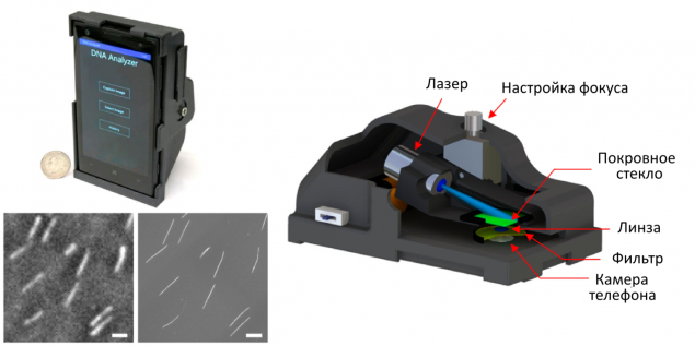
Image description: appearance are running a mobile application scheme of its internal structure and pictures of DNA fragments left - obtained by the proposed devaysa right - using a fluorescence microscope. Scale - 10 micrometers.
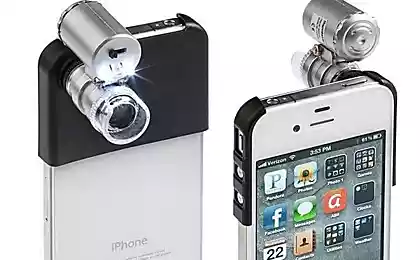
The design of the device is relatively simple: a mobile phone with built-in camera (in this study used a smartphone Lumia 1020) equipped with an additional magnifying lens seized from another phone, and a laser with a wavelength of 450 nm. Laser excitation was necessary for the fluorescence dye which labeled DNA fragments studied. The sample was placed between two bits of glass and lit his laser, with the dye emits light, which fixed camera phone. Additional filters are placed between the two lenses, passed a glow dye and detain any non-specific signal. Total sample was done 10-15 shots, each - delayed about 4 seconds. The obtained data were processed using a special algorithm that calculates the length of the DNA fragments caught in the frame. It is assumed that these calculations will be made not on the phone, and on a dedicated server: mobile application will send images to the processing and return the results to the user ready.
The total weight of the device is about 190 grams, including three AAA batteries to power the laser, and its cost, the assurances of the authors, not more than 400 euros. At the same time obtained using this photograph of a simple device, though inferior in quality to images taken with a conventional fluorescence microscope, but allow to cover a large area of the shooting - about 2 mm2. At the same time manage to get images of the DNA fragments in the size of 10 kb and above (1 kilobase = 1000 nucleotide base pairs), and developed software allows to achieve acceptable accuracy in the measurement of the length of the fragments (average error - less than 1 kb), and the measurement accuracy increases with the length fragments.
The authors believe that their invention can be extremely useful for the rapid diagnosis of cancer or neurological diseases associated with changes in the length of individual sections of the genome. In addition, the authors hope that their work will encourage other researchers to search for innovative ways to use modern mobile technology - smartphones and tablet computers.
Source: geektimes.ru/post/243727/
Access to Gmail in China partially restored
Airline sued the owner of a site that offers discount tickets to the unexpected
