3620
The logic of thinking. Part 1. Neuron
Year and a half ago, I put on the Habr cycle video lectures with my vision of how the brain works and what are the possible ways to create artificial intelligence. Over the past since the time managed to move forward. Something happened a deeper understanding that somehow managed to simulate on a computer. What is nice, there were supporters, actively participating in the project.
In this series of articles planned to talk about the concept of the intelligence on which we are working and demonstrate some of the decisions that are fundamentally new in the field of modeling of the brain. But that story was clear and consistent, it will contain not only a description of new ideas, but also the story of how the brain works in general. Some things, especially at the beginning may seem simple and well-known, but I would advise you not to miss them, since they largely determine the overall conclusiveness of the narrative.
The general idea of the brain
Nerve cells, they also neurons, with their fibers transmitting signals form the nervous system. In vertebrates, the bulk of the neurons are concentrated in the cranial cavity and the spinal canal. This is called the central nervous system. Accordingly, the isolated brain and spinal cord as its components.
Spinal cord collects signals from most receptors body and transmits them to the brain. Through the structure of the thalamus they are distributed and projected onto the cerebral cortex of the brain.
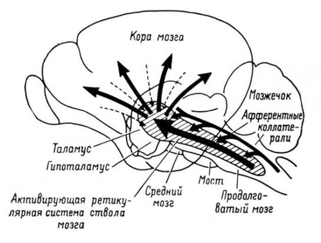
The projection information on the bark i>
In addition to the cerebral hemispheres is engaged in information processing is also the cerebellum, which is, in fact, is a small independent brain. Cerebellum provides accurate motor skills and coordination of all movements.
Vision, hearing and smell brain provide the flow of information about the outside world. Each of the components of this stream, passing on its path, and is projected onto the crust. Bark - a thick layer of gray matter from 1.3 to 4.5 mm and constituting an outer surface of the brain. Due to the convolutions formed folds, bark packed so that it takes three times less space than in the expanded form. The total area of the cortex of one hemisphere - about 7,000 square centimeters
As a result, all signals are projected onto the crust. The projection is carried bundles of nerve fibers, which are distributed on a limited area of the cortex. The plot, which is projected or external information, or information from other parts of the brain forms a zone of the cortex. Depending on which signals arrive at such a zone, it has its specialization. Distinguish the motor area of the cortex, sensory area, Broca's area, Wernicke's visual areas, occipital lobe, only about a hundred different areas.
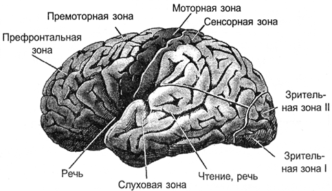
cortex i>
In the vertical direction the cortex can be divided into six sections. These layers do not have clear boundaries and are defined by the prevalence of a particular cell type. In various cortical areas these layers can be expressed in different ways, more or less. But, in general, we can say that the crust is flexible enough, and assume that the functioning of its various zones is subject to the same principles.
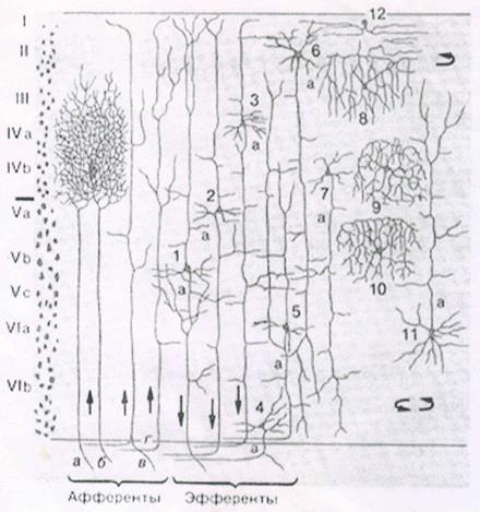
Layers of bark i>
By afferent fibers signals are sent to the cortex. They fall on the III, IV level of the cortex where spread to nearby to the place where it fell afferent fiber neurons. Most neurons have axonal connections within their area of the cortex. But some neurons have axons that go beyond it. For these efferent fibers signals are either outside the brain, such as the executive bodies, or projected onto other parts of the cortex of his or the other hemisphere. Depending on the direction of signal transmission efferent fibers can be divided into:
associative fibers that connect the individual parts of the cortex of one hemisphere; commissural fibers that connect the two hemispheres of the cortex; projection fibers that connect the cortex with the nuclei of the lower parts the central nervous system.
One can imagine the cerebral cortex as a great sheet, cut out for individual zones. Picture of neuronal activity of each zone encodes certain information. Bundles of nerve fibers formed by axons that go beyond its cortex, forming a system of projection links. At each of the zones is projected certain information. And one area can enter multiple information streams that can come with both of his bands, and the opposite hemisphere. Each flow of information similar to the kind of picture drawn activity axons nerve bundle. Functioning separate cortex - is providing a plurality of projections, memorization of information, its processing, the formation of his own painting activity and further projection information, resulting in a work of this zone.
A significant volume of the brain - a white substance. It is formed by the axons of neurons, creating the very flat way. In the figure below you can see the white matter how bright the filling between the cortex and the internal structure of the brain.
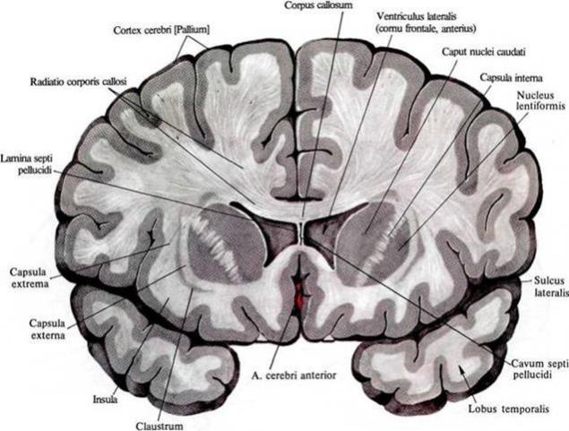
The distribution of white matter in the frontal sections of the brain i>
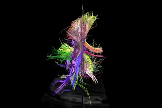
Using spectral diffuse MRI was able to track the direction of the individual fibers and build a three-dimensional model of connectedness cortical areas (project Connectomics (CONNECT)).
Understanding of the structure of relations is well below the figures given (Van J. Wedeen, Douglas L. Rosene, Ruopeng Wang, Guangping Dai, Farzad Mortazavi, Patric Hagmann, Jon H. Kaas, Wen-Yih I. Tseng, 2012).
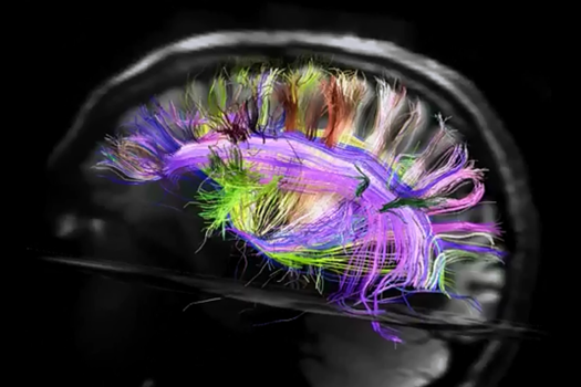
View from the left hemisphere i>
Rear view i>
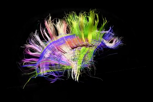
Right view i>
By the way, a rear projection is clearly visible asymmetry tract of the left and right hemispheres. This asymmetry is largely determines the differences in those functions that acquire hemisphere as they are learning.
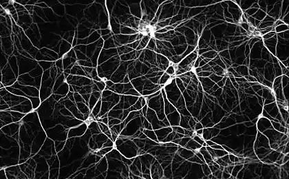
Neuron
Base of the brain - neuron. Naturally, the modeling of the brain using neural networks begins with the answer to the question, what is the principle of its operation.
At the heart of a real neuron are chemical processes. In the resting state between the inside and the outside of the neuron, there is a potential difference - the membrane potential of around 75 mV. It is formed by the operation of specific protein molecules that act as sodium-potassium pump. These pumps due to the energy nucleotide ATP driven potassium ions inside and sodium ions - out of the cell. Since the protein thus acts as an ATP-ase, i.e. the enzyme hydrolyzing ATP, it is called - "sodium-potassium ATPase." As a result, the neuron is converted into a charged capacitor with a negative charge inside and outside positive.
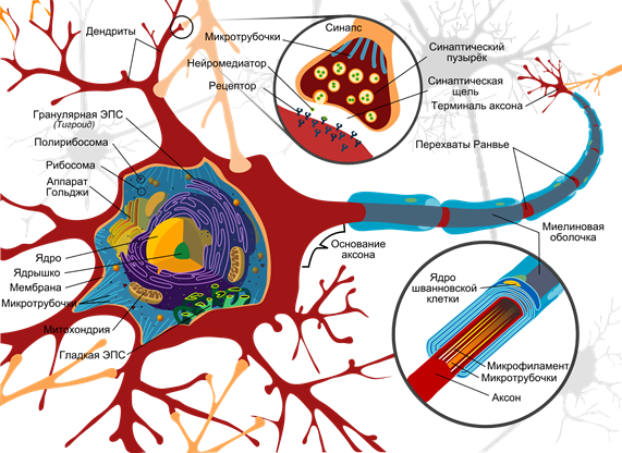
The circuit neuron (Mariana Ruiz Villarreal) i>
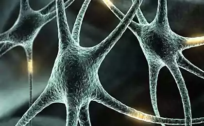
The surface is covered with a neuron branching processes - dendrites. By the end of the axon adjacent dendrites of other neurons. Their joints are called synapses. Through the interaction of synaptic neuron can respond to the incoming signals and under certain circumstances to generate its own momentum, called spike.
Signal Transmission at synapses happens through chemicals called neurotransmitters. When a nerve impulse along the axon enters the synapse, it releases molecules of neurotransmitter vesicles special characteristic of this synapse. On the membrane of a neuron, the receiving signal has a protein molecule - receptors. Receptors interact with neurotransmitters.
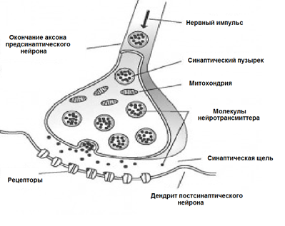
Chemical synapse i>
Receptors located in the synaptic cleft are ionotropic. It emphasizes the fact that they are the same ion channels, capable of moving ions. Neurotransmitters act on the receptors so that their ion channels open. Accordingly, a membrane is depolarized, hyperpolarizing or - depending on which channels are affected, and, accordingly, this type of synapse. At excitatory synapses open channels permeable cations into the cell - membrane is depolarized. In inhibitory synapses open channels, providing anions, leading to membrane hyperpolarization.
In certain circumstances, the synapses can change their sensitivity is called synaptic plasticity. This leads to the fact that one neuron synapses become different between a susceptibility to external signals.
Simultaneously, the synapses of a neuron receives a plurality of signals. Inhibitory synapses pull the membrane potential toward the accumulation of charge inside the cage. Activating synapses, on the contrary, trying to defuse the neuron (shown below).
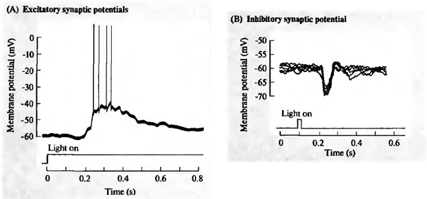
Excitation (A) and inhibition (B) retinal ganglion cells (J. Nicholls., Martin R., Wallace B., P. Fuchs, 2003) i>
When the total activity exceeds the threshold of initiation, a discharge, called an action potential or spike. Spike - a sharp depolarization of the membrane of a neuron, which generates an electrical impulse. The entire process of generating momentum lasts about 1 millisecond. However, neither the length nor the amplitude of the pulse does not depend on how strong were the reasons advanced (see Figure below).
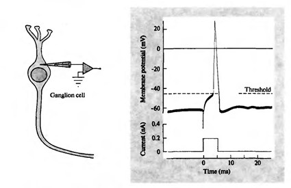
Join the action potential ganglion cells (J. Nicholls., Martin R., Wallace B., P. Fuchs, 2003) i>
After Spike ion pumps provide the reuptake of the neurotransmitter and clearing the synaptic cleft. During the refractory period, coming after the spike, the neuron is not able to generate new impulses. This period determines the maximum oscillation frequency, which is capable of neuron.
Spikes that arise as a result of activity in the synapses, called induced. The repetition frequency of spikes caused encodes how well the input signal corresponds to setting the sensitivity of the neuron synapses. When the signals falls on sensitive synapses activate neurons, and it does not interfere with the signals arriving at inhibitory synapses, the response of the neuron is maximized. The image that is described by the signals characteristic of the neuron is called a stimulus.
Of course, the idea of the neurons is not necessary to oversimplify. Information between certain neurons can be transmitted not only spikes, but also by connecting channels intracellular contents and transmitting the electrical potential directly. Such an extension is called gradual, but the compound itself is called an electrical synapse. Dendrites, depending on the distance from the cell body are divided into proximal (close) and distal (remote). The distal section can form dendrites, which act as semi-elements. In addition to synaptic pathways have extrasynaptic excitation mechanisms causing metabotropic adhesions. In addition there is also evoked activity and spontaneous activity. Finally, the neurons of the brain are surrounded by glial cells, which also have a significant impact on the running processes.
The long road of evolution has created many mechanisms used by the brain in their work. Some of them can be understood by themselves, meaning other becomes clear when considering only the fairly complex interactions. So do not take the above description is made of a neuron as exhaustive. To go to the deeper patterns, we must first deal with the "basic" properties of neurons.
In 1952, Alan Lloyd Hodgkin and Andrew Huxley were made describing the electrical mechanisms that determine the generation and transmission of nerve signals in the squid giant axon (Hodgkin, 1952). What was rated the Nobel Prize in Physiology or Medicine in 1963. Model Hodgkin - Huxley describes the behavior of a neuron system of ordinary differential equations. These equations correspond to the self-oscillatory processes in the active medium. They take into account the set of components, each of which has its counterpart in the real biophysical cell (figure below). Ion pumps correspond to a current source I p sub>. The inner lipid layer of the cell membrane forms a capacitor with capacitance C m sub>. Ion channels synaptic receptors provide electrical conductivity g n sub>, which depends on the input signals change with time t, and the total value of the membrane potential V. The leakage current of membrane pores creates conductor g L sub> . The movement of ions on ion channels occurs under the influence of electrochemical gradients, corresponding to a voltage source with an electromotive force E n sub> and E L sub>.
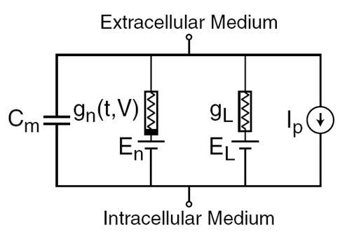
The main components of the model of Hodgkin - Huxley i>
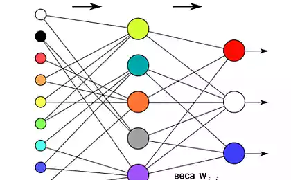
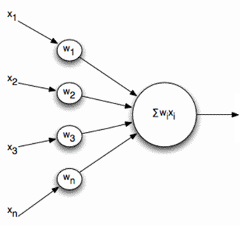
Naturally, with the creation of neural networks have a desire to simplify the model of a neuron, leaving only the most essential properties. The most famous and popular simplified model - an artificial neuron McCulloch - Pitts, developed in the early 1940s (J. McCulloch., W. Pitts, 1956).
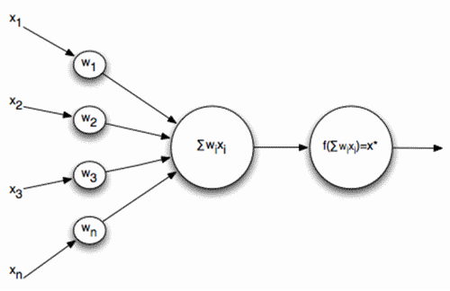
The formal neuron McCulloch - Pitts i>
The inputs of the neuron signals are. These signals are summed balanced. Next to this linear combination used some non-linear activation function, for example, a sigmoid. Often used as a sigmoidal logistic function:
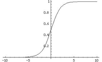
The logistic function i>
In this case the activity is recorded as a formal neuron
As a result of such a neuron becomes a threshold adder. At sufficiently steep threshold function of the neuron output signal - either 0 or 1. A weighted sum of the input signal and the weights of the neuron - a convolution of two images: the image of the input signal and the image described by the weights of the neuron. The result of the convolution of the higher, the more accurate matching of these images. That is, the neuron is in fact determines how the signal is similar to the supplied image, recorded at its synapses. When the value of the convolution exceeds a certain level and the threshold function is switched to one, it can be interpreted as a strong statement of the neuron that he learned presenting the image.
Real neurons are indeed somehow similar to neurons McCulloch - Pitts. The amplitudes of the spikes is not dependent on what signals they were called synapses. Spike, either there or it is not. But the real neurons respond to a stimulus not a single pulse and pulse sequence. The frequency of the pulse, the higher know exactly typical neuron image. This means that if we construct a neural network of adders such thresholds, it is in a static input signal and provide at the output a result, but the result will be far from the reproduction of how the actual neurons. In order to bring the neural network to the biological prototype, we need to simulate the dynamics of a given timing and frequency properties of reproducing signals.
But you can go the other way. For example, you can select the generalized characteristic of neural activity, which corresponds to the frequency of its pulses, ie the number of spikes over a certain period of time. If you go to this description, one can imagine a neuron as a simple linear combiner.
linear combiner i>
And output signals, respectively, for the input of such neurons are not dihatomichnymi (0 or 1), and expressed in some scalar quantity. The activation function of time is recorded as

Linear adder should not be taken as something fundamentally different compared with pulsed neuron, it just allows for modeling or description go to longer time intervals. Although pulsed description more correctly, the transition to a linear combiner is justified in many cases a strong simplification of the model. Furthermore, some important properties are difficult to discern in a pulsed neuron readily apparent to the linear combiner.
Continued
References
Alex Redozubov (2014)
Source: habrahabr.ru/post/214109/