1961
The winners
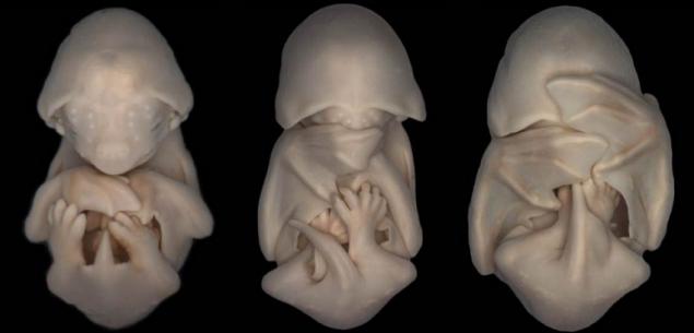
Typically, all pictures taken with different magnification and using different techniques of shooting. But the fact that they allow you to see the mysterious world, the existence of which most people do not realize is admired. 20th place. Bright field micrograph of three embryos black Molos (species of bats) in the progressive stage of development. A little later, their wings begin to lengthen and ears - to become more
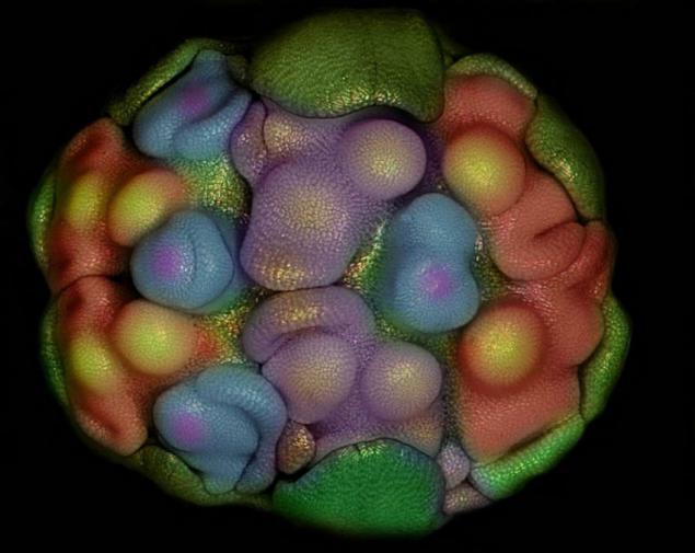
19th place. Cell rudiments (primordia), is the earliest stage of development of the flower. The photo was taken with the illumination technique of Kirlian effect
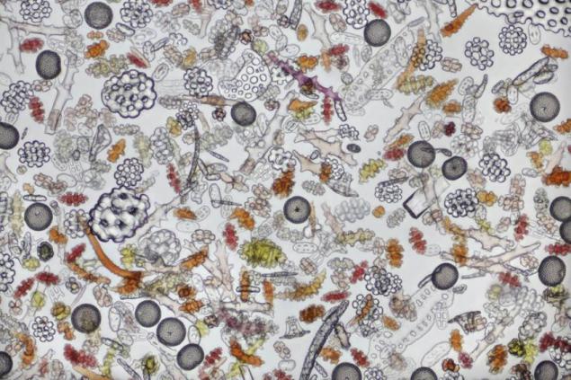
18th place. Bright field micrograph of particles of various forms of coral sand. 100-fold increase
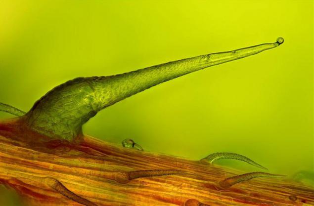
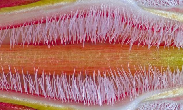
17th place. Stinging nettle leaves at the tip. Micrograph made using the technique of the transmitted light. 100-fold increase
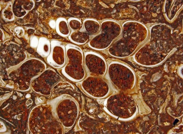
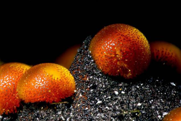
16th place. The cut shell fossil turitelly containing ancient freshwater snails and ostracods. 7-fold increase
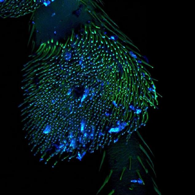
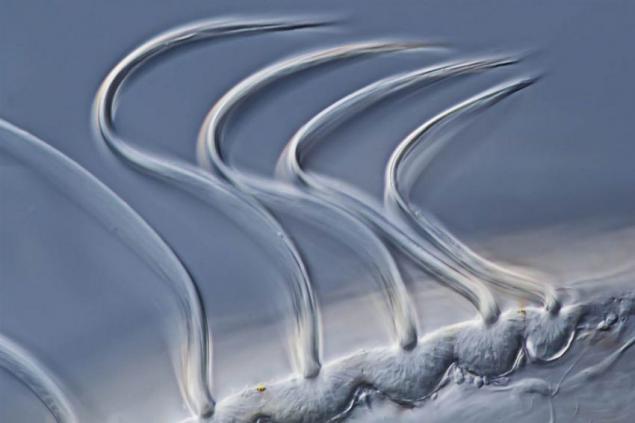
15th place. Confocal image of the foot ladybug. 10-fold increase
14th place. Pink, orange and yellow colors abound on the pistil of fat Adenium (desert rose). Photo made in the technique giperfokusirovki. 10-fold increase
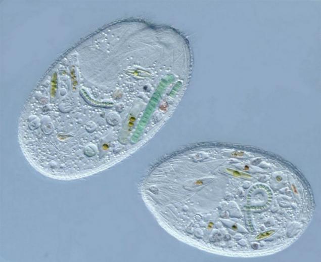
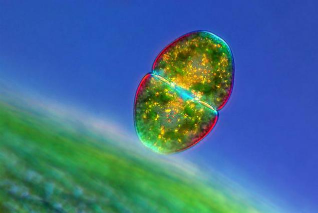
13th place. Type the simplest known as Sonder. These ciliates love to feast on cyanobacteria and diatoms. 400-fold increase
12th place. Confocal fluorescence image, and lymphatic endothelial cells and fibroblasts. 200-fold increase
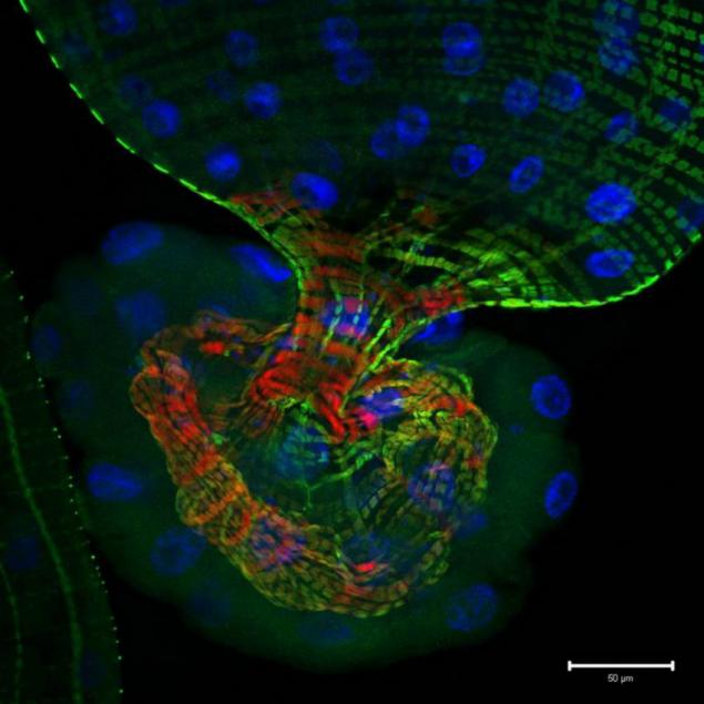
11th place. Confocal image of part of the intestine of the fruit fly larvae that contains markers signaling activity Notch (green), the cytoskeleton (red) and nuclei (blue). 25-fold increase.
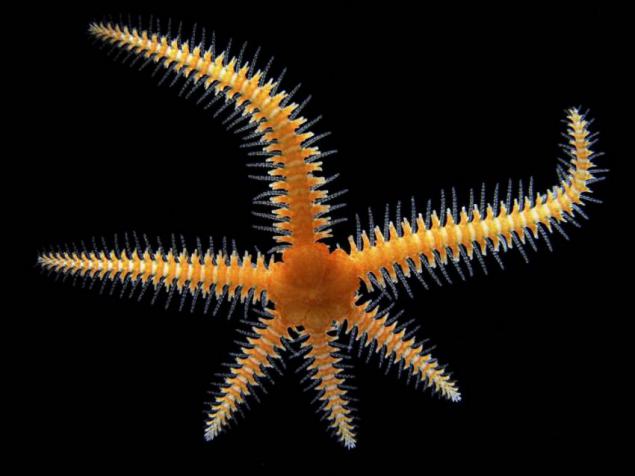
10th place. Stereomicroscopical and dark-field image zmeehvostki. 8-fold increase
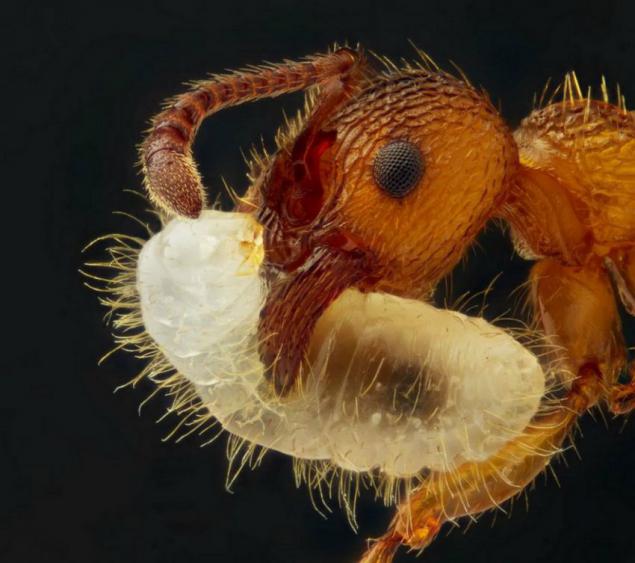
9th place. Ant carrying a larva. The photo was taken giperfokusirovki technique and reflected light. 5-fold increase.
8th place. Micrograph larvae plevrobrahii, made in the technique of differential interference contrast. 500-fold increase
7th place. Confocal image of the eye of the fruit fly. 60-fold increase
6th place. COSMAR (seaweed) near the leaves sfagnida (moss) in polarized light. 100-fold increase
5th place. Micrograph of tiny spheres of mine mineral kakoksena La Paloma, in the technique of the transmitted light. 18-fold increase.
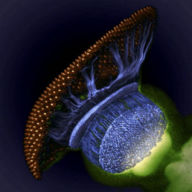
4th place. Confocal image of the visual system of the fruit fly pupa stage of development. The retina is a golden color, the photoreceptors axons - blue, brain tissue - green. 1500-fold increase.
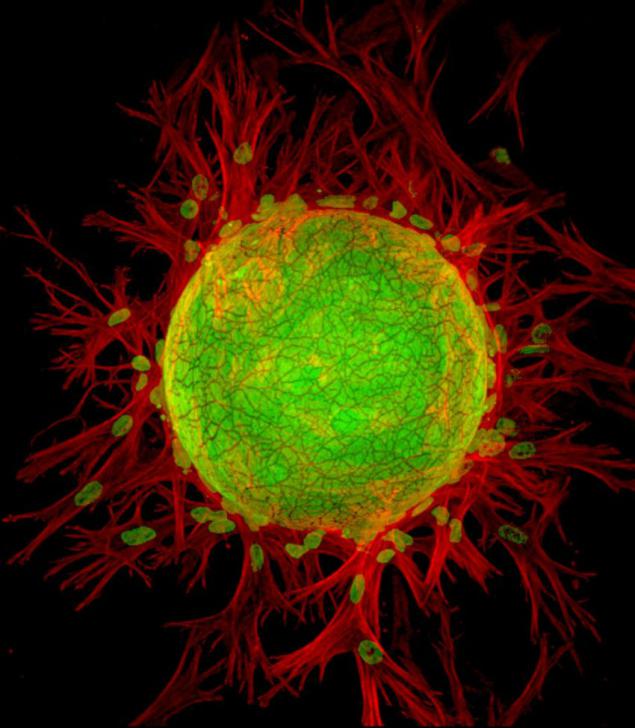
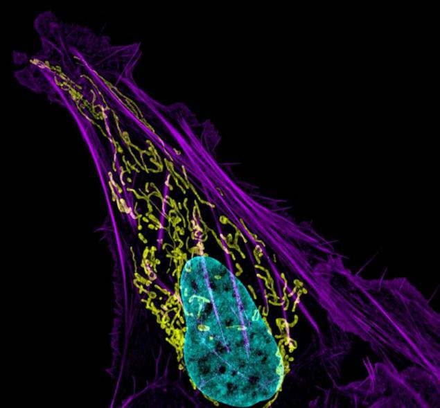
3rd place. Human bone cancer. Actin fibers are shown in purple, mitochondria - yellow, DNA - blue. 63-fold image.
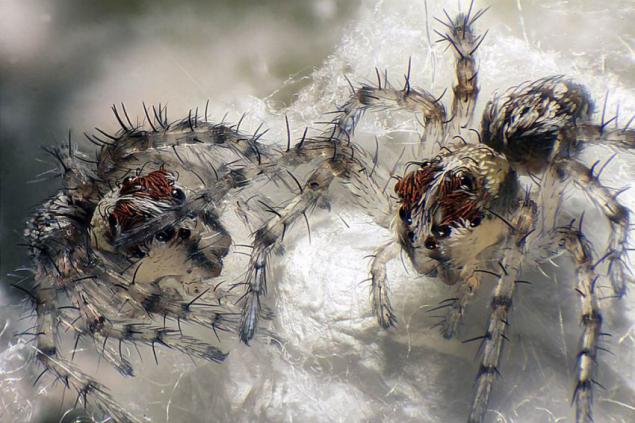
2nd place. Micrograph of newborn lynx spiders, in the technique of the reflected light, fiber optics and giperfokusirovki. 6-fold increase
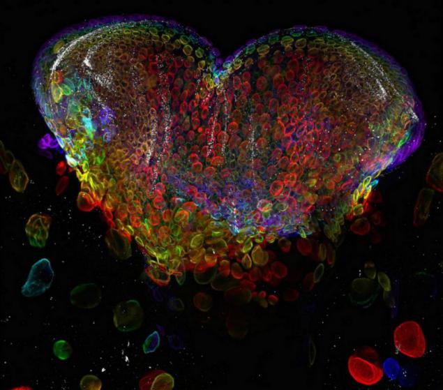
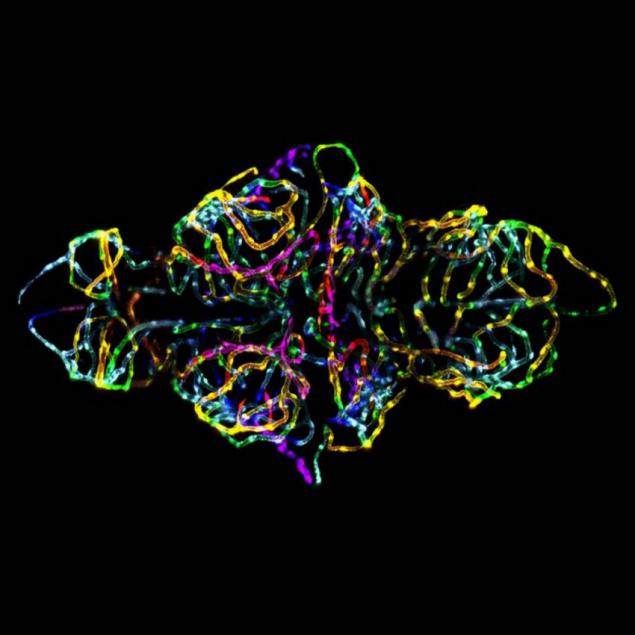
1st place. Confocal image of the blood-brain barrier in zebrafish embryos. Photo is notable because it is the first image of the blood-brain barrier is a living creature. 20-fold increase
Source: jurashz.livejournal.com

19th place. Cell rudiments (primordia), is the earliest stage of development of the flower. The photo was taken with the illumination technique of Kirlian effect

18th place. Bright field micrograph of particles of various forms of coral sand. 100-fold increase

17th place. Stinging nettle leaves at the tip. Micrograph made using the technique of the transmitted light. 100-fold increase

16th place. The cut shell fossil turitelly containing ancient freshwater snails and ostracods. 7-fold increase

15th place. Confocal image of the foot ladybug. 10-fold increase

14th place. Pink, orange and yellow colors abound on the pistil of fat Adenium (desert rose). Photo made in the technique giperfokusirovki. 10-fold increase

13th place. Type the simplest known as Sonder. These ciliates love to feast on cyanobacteria and diatoms. 400-fold increase

12th place. Confocal fluorescence image, and lymphatic endothelial cells and fibroblasts. 200-fold increase

11th place. Confocal image of part of the intestine of the fruit fly larvae that contains markers signaling activity Notch (green), the cytoskeleton (red) and nuclei (blue). 25-fold increase.

10th place. Stereomicroscopical and dark-field image zmeehvostki. 8-fold increase

9th place. Ant carrying a larva. The photo was taken giperfokusirovki technique and reflected light. 5-fold increase.

8th place. Micrograph larvae plevrobrahii, made in the technique of differential interference contrast. 500-fold increase

7th place. Confocal image of the eye of the fruit fly. 60-fold increase

6th place. COSMAR (seaweed) near the leaves sfagnida (moss) in polarized light. 100-fold increase

5th place. Micrograph of tiny spheres of mine mineral kakoksena La Paloma, in the technique of the transmitted light. 18-fold increase.

4th place. Confocal image of the visual system of the fruit fly pupa stage of development. The retina is a golden color, the photoreceptors axons - blue, brain tissue - green. 1500-fold increase.

3rd place. Human bone cancer. Actin fibers are shown in purple, mitochondria - yellow, DNA - blue. 63-fold image.

2nd place. Micrograph of newborn lynx spiders, in the technique of the reflected light, fiber optics and giperfokusirovki. 6-fold increase

1st place. Confocal image of the blood-brain barrier in zebrafish embryos. Photo is notable because it is the first image of the blood-brain barrier is a living creature. 20-fold increase

Source: jurashz.livejournal.com