2358
The world under the microscope: the best picture in 40 years
Colonial plankton (250-fold increase), Wim van Egmond, 2013.
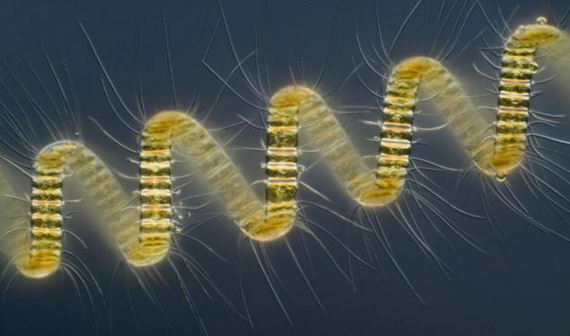
The spread of the most diverse - from increased 250 times colonial plankton head to fly under 6, 25-fold zoom. It is impossible to count how many were sent to work at Small World Photomicrography Competition in history, but a rare year in the last 10 years go by without a half to two thousand candidates.
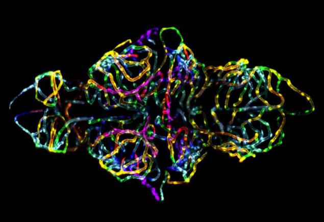
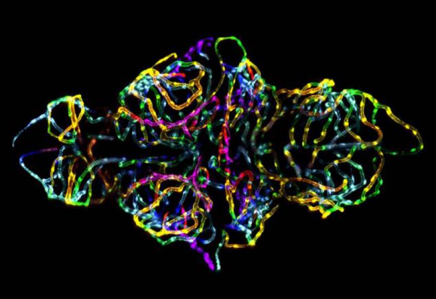
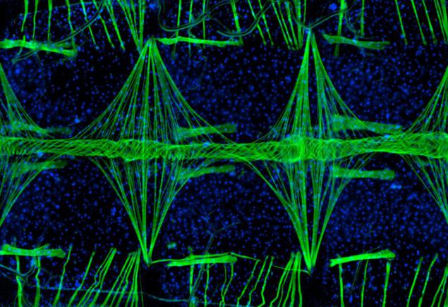
The blood-brain barrier (20X), Jennifer L. Peters and Michael R. Taylor, 2012.
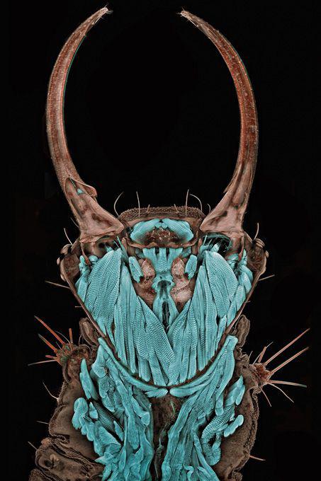
Portrait of a green lacewing larva (20x), Igor Sivanovich, 2011.
Heart tissue mosquito (100x), Jonas King, 2010.
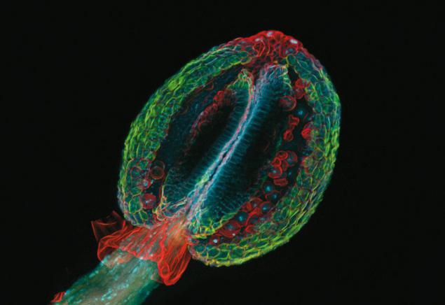
Pestle Arabidopsis thaliana (20X), Tallinn Technical University, 2009.
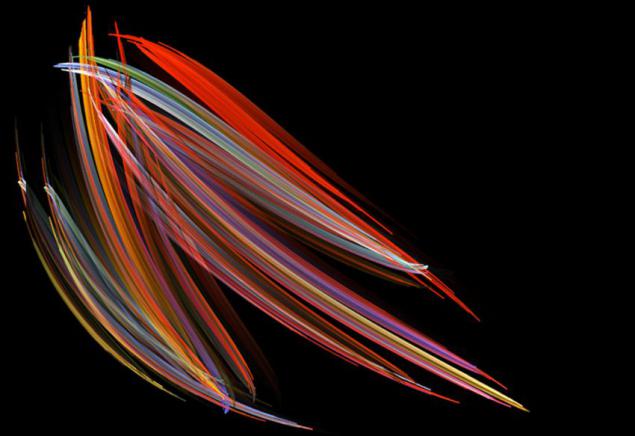
Marine diatoms (200x), Michael Stringer, 2008.
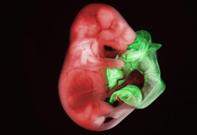
Double transgenic mouse embryo (17x), Gloria Kwon, 2007.
Mouse colon (740h), Paul L. Appleton, 2006.
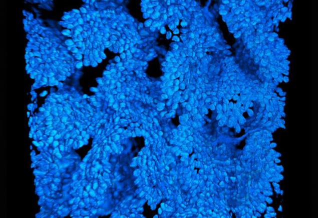
The head flies (6, 25x), Charles B. Krebs, 2005.
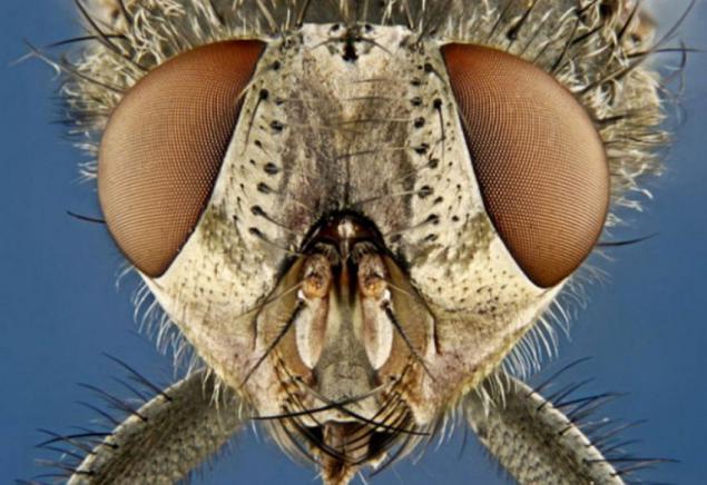
Quantum dots are nanocrystals deposited on a silicon substrate (200x), Seth A. Coe Sullivan, 2004.
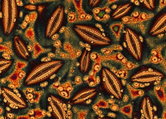
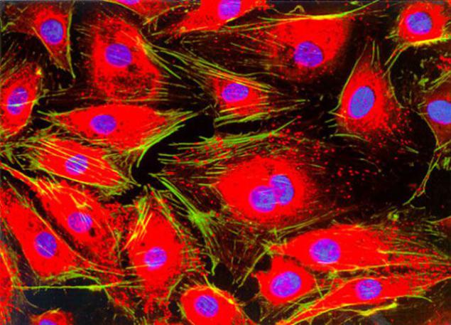
Structural proteins in the cells of mice (100x), Torsten Wittmann, 2003.
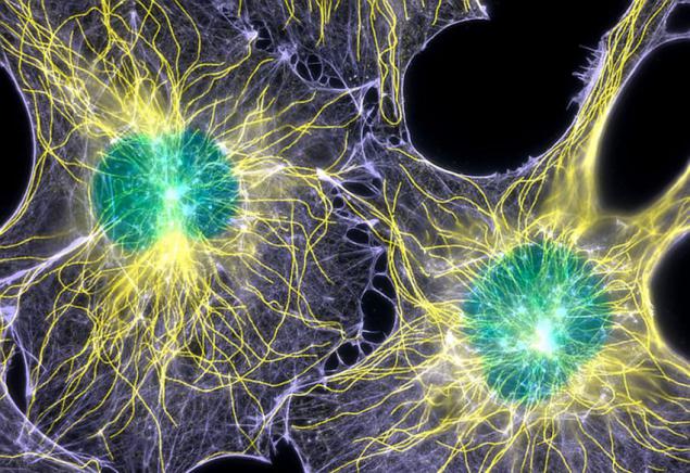
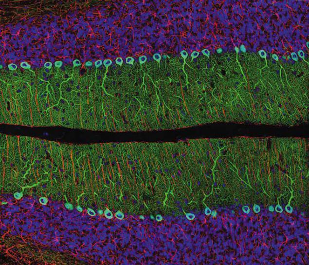
Sagittal section of rat cerebellum (40x), Thomas J.. Derinkuyu, 2002.
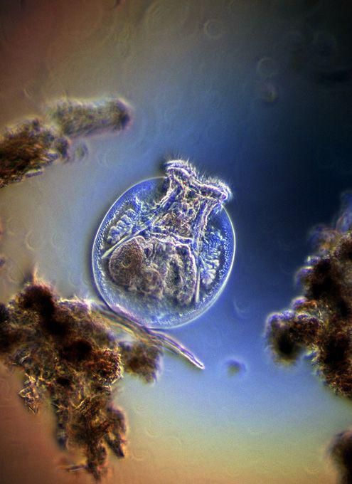
Rotifers in freshwater (200x), Harold Taylor Kensvort, 2001.
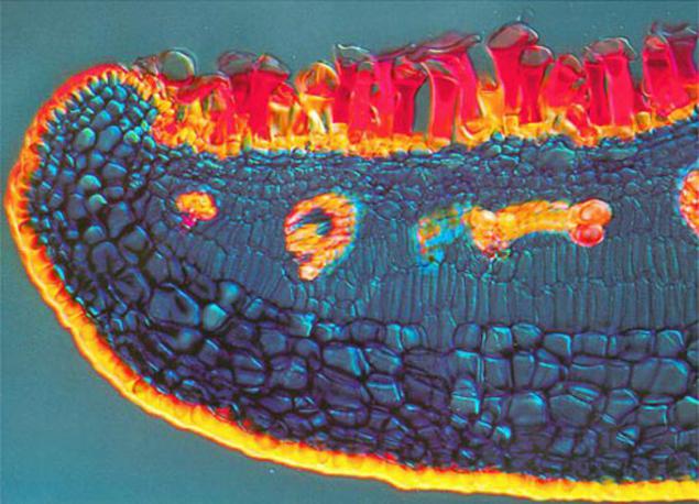
Avicennia marina (40x), Daphne Zbayren Colborne, 2000.
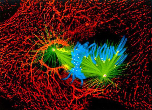
Lung cells in mitosis (240x), Alexei Khodzhakov, 1999.
Endothelial cells (100x), Jakob Zbaeren, 1998.
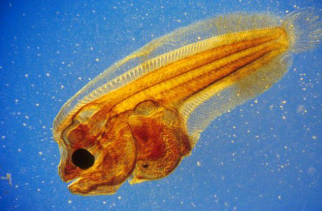
Larva Pleuronectidae (20x), Christian Gautier, 1995.
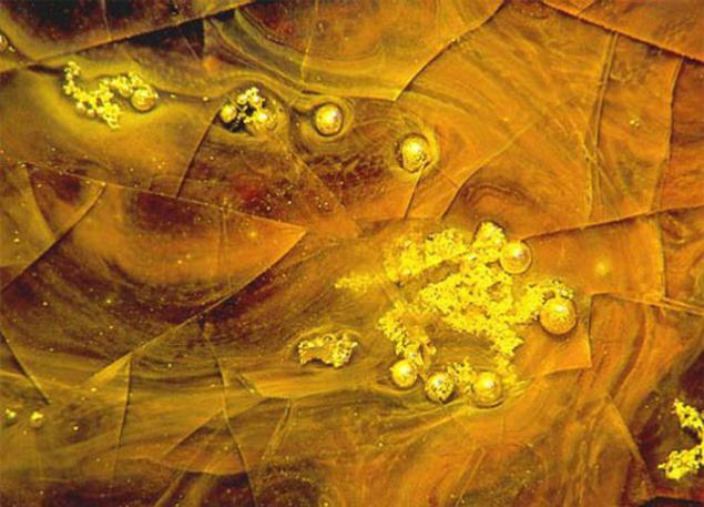
Gold deposit in the glassy matrix (20x), David Smith, 1988.
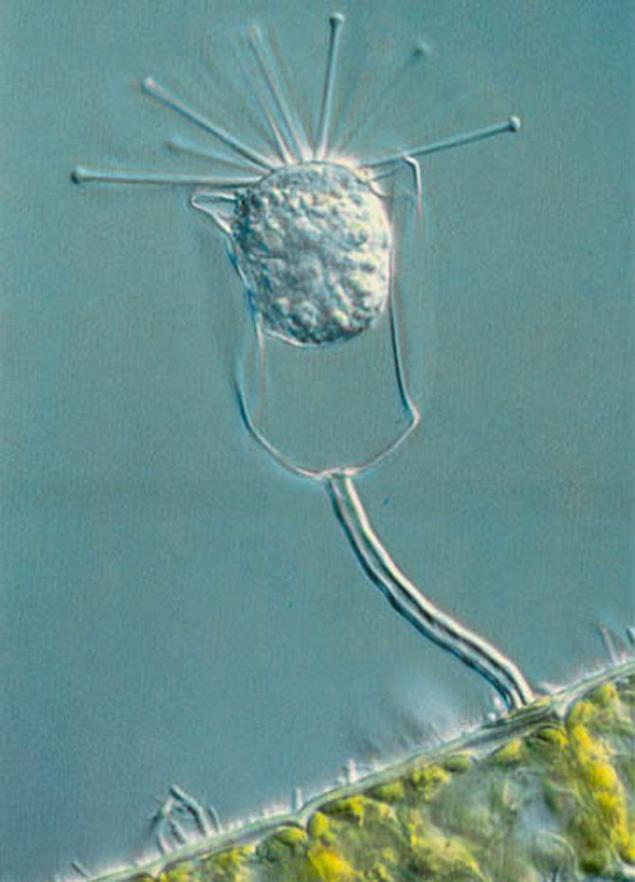
The simplest filamentous algae with bacteria on the surface (160X), Paul W. Johnson, 1979.
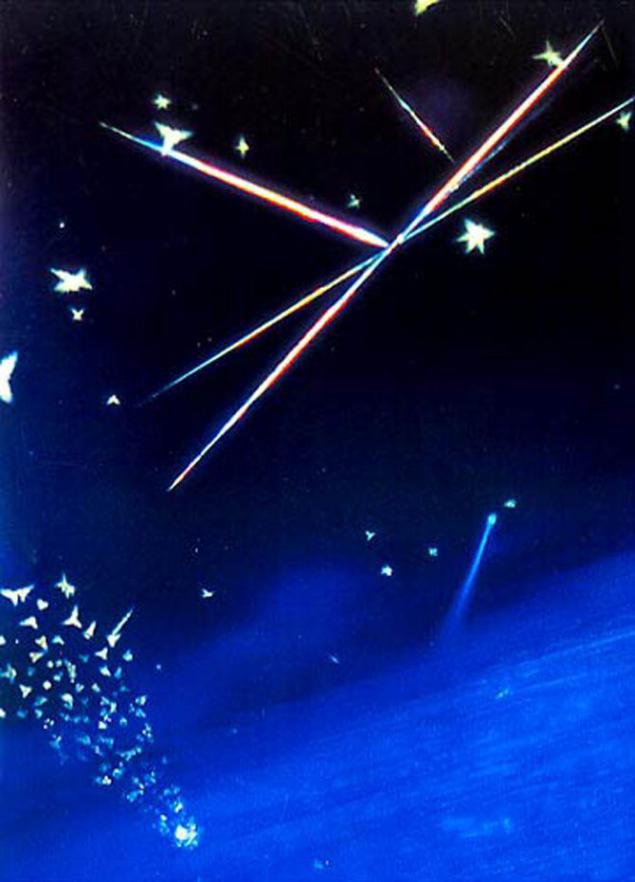
Crystals of rutile and tridymite in glass, cobalt enriched (350x), James W. Smith, 1977.
Source: trendymen.ru