2897
Photos from the competition micrographs of Nikon (19 photos)
International Competition micrographs of Nikon's recently announced the winners of its 2010. The contest was founded in 1974 specifically for those who like to take pictures through a microscope. Looking to the world of animals, plants and minerals with the help of new technologies and the various tools contestants this year, we offer photos of crystalline formations, fluorescent body parts, reticulated structure and more of value because of its beauty and inner peace.
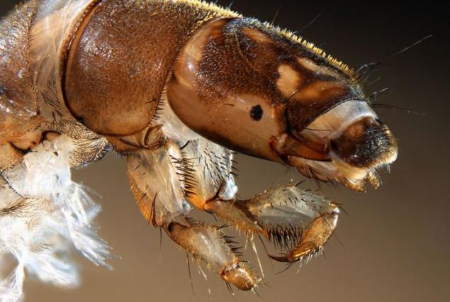
Increased 30-fold image of the head caddis larvae made Fabrice couple from Caen, France. (Courtesy of Nikon Small World)
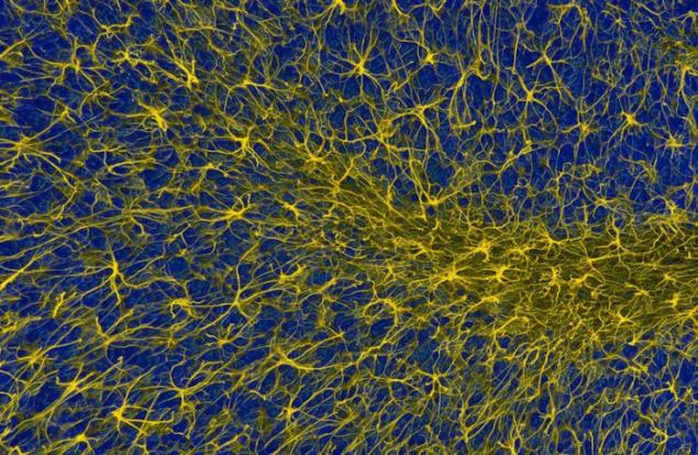
Magnified 400 times fluorescent photos of glial cells in the cerebellum. Glial cells provide support to neurons in the brain. The photo was taken by Thomas Deerinkom from the National Center for Microscopy at the University of California, San Diego. (Courtesy of Nikon Small World)
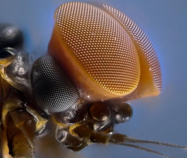
Volchkoobraznye eyes of the male fly-by-night. Photo enlarged 10 times. Author - Laurie Knight of Konbridzha, Kent, England. (Courtesy of Nikon Small World)
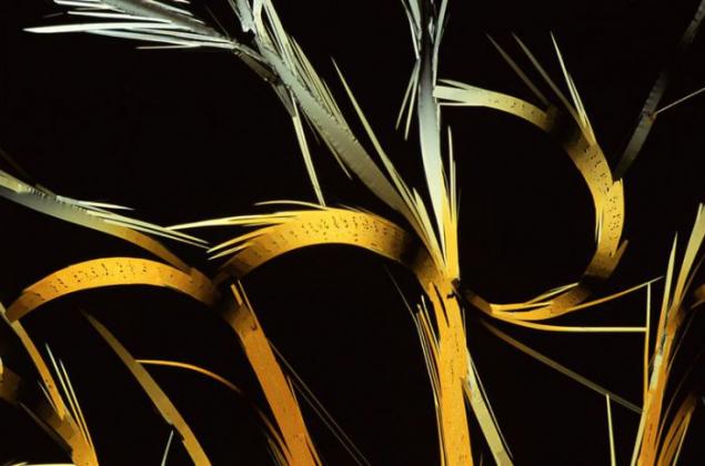
Increased 63 times Photo recrystallized sulfur made by Dr. Edward Leimane Gaffordom of Ventura, California. (Courtesy of Nikon Small World)
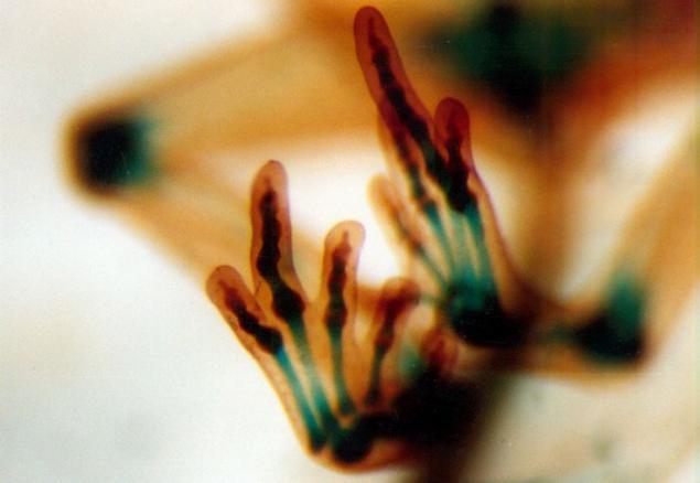
Tiny bones in the limbs developing frog. The image is enlarged 20 times. Author - Dr. Mike Klimovskaya from the University of Colorado at Boulder. (Courtesy of Nikon Small World)
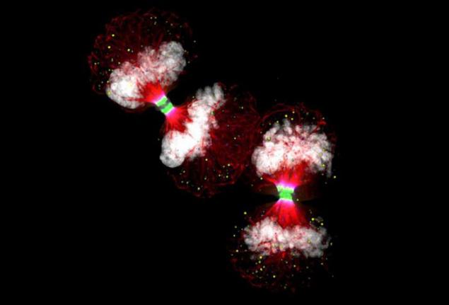
Two human cancer cells before division by four. Image is enlarged 100 times. It won 11 seats and was made by Dr. Paul D. Andrews of the University of Dundee, Scotland. (Courtesy of Nikon Small World)
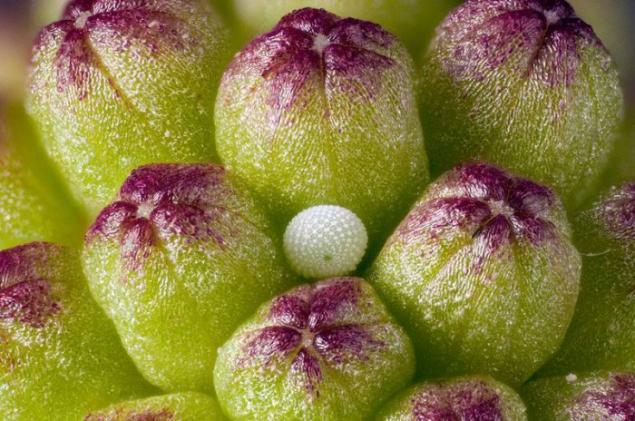
Egg Green hairstreak in pink rose buds. The picture is enlarged six times. It was made by David Millard from Austin, Texas. (Courtesy of Nikon Small World)
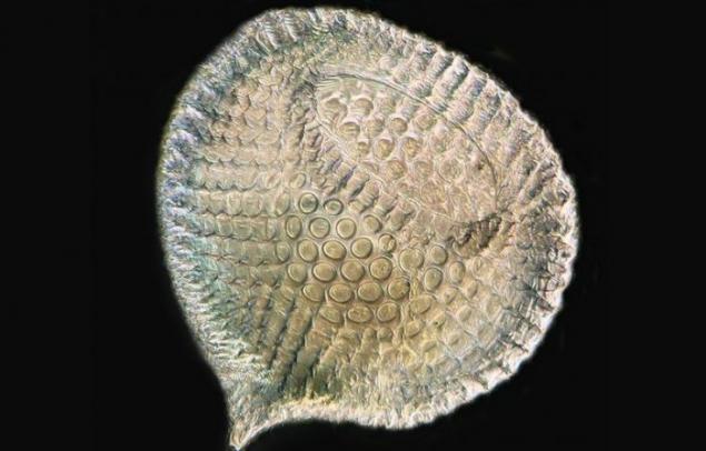
Radiolarian at a magnification of 250 times. The picture was taken by Raymond Sloss of the Historical Society in Northampton, UK. (Courtesy of Nikon Small World)
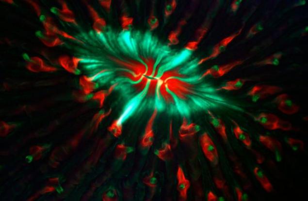
James Nicholson of the Research Institute in Charleston, South Carolina, Fung took this picture with natural fluorescent proteins around the mouth. Snapshot increased by 6 times. (Courtesy of Nikon Small World)

Magnified 250 times Photo of red algae from Dr. Arlene Vechezak from Anacortes, WA. (Courtesy of Nikon Small World)
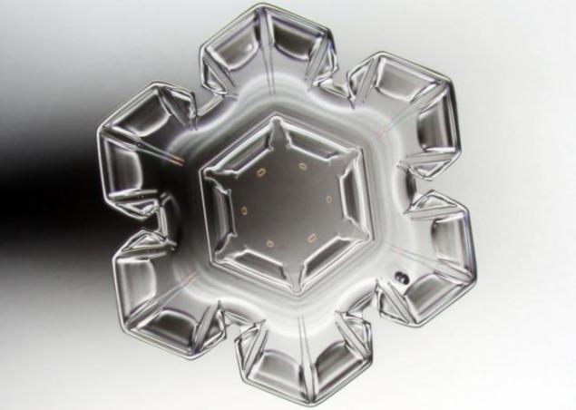
Pekka Honkakoski of Sonkajärvi, Finland, took this picture of the crystal snowflakes, increased 40 times. (Courtesy of Nikon Small World)
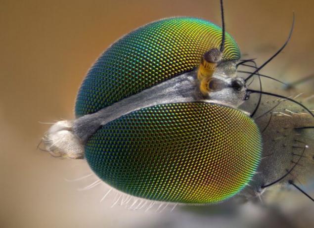
Increased 10-fold photo flies made by Laurie Knight of Tonbridge, Kent, England. (Courtesy of Nikon Small World)
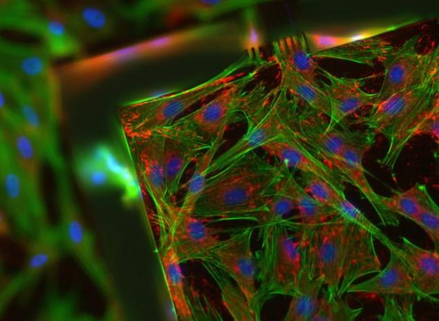
Subcutaneous rat cells. The picture is enlarged 20 times and made Rafael Pennes from Lausanne, Switzerland. (Courtesy of Nikon Small World)
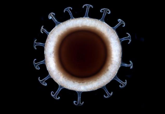
Pearlwort at a magnification of 20 times. The picture was taken Jocelyn Cheng of Rochester Institute of Technology. (Courtesy of Nikon Small World)
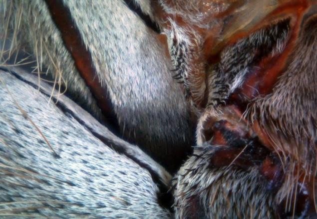
Segments paws tarantulas. The picture is enlarged 40 times and made Tireli Pinnegarom from Nanaimo, Canada. (Courtesy of Nikon Small World)
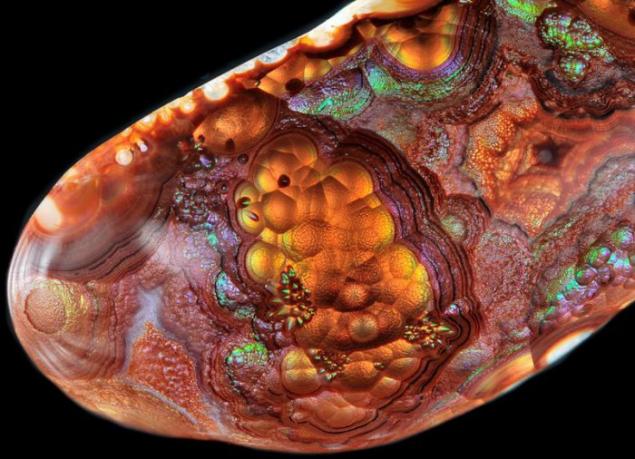
Mexican agate polished piece at a magnification of 4 times. Author - Thomas Shearer of Duluth, Minnesota. (Courtesy of Nikon Small World)
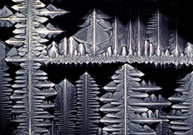
Crystals geksatsianozhelezo-acid potassium. The picture is enlarged 40 times and made Stefan Eberhard of the University of Georgia. (Courtesy of Nikon Small World)
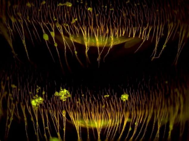
Increased 40 times shot belly bees with pollen particles. Author - Dr. Robert Markus Institute of Genetics in the biological center of the Hungarian Academy of Sciences in Szeged. (Courtesy of Nikon Small World)
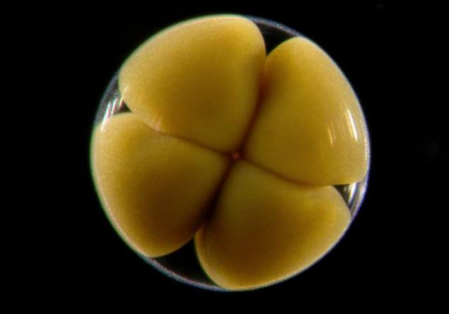
Embryo starfish on the fourth stage of the cell at a magnification of 60 times. The picture was taken by Dr. Alvaro Migotto of the University of Sao Paulo, Brazil. (Courtesy of Nikon Small World)