1423
Museum of Medical History (40 photos)
A look at some monstrous exhibits difficult. It is heartbreaking sight, and the viewer is experiencing a real psychological shock. But the picture of anomalies of the human body is extremely attractive from an intellectual standpoint. Because it gives you the answer to one of the oldest questions facing man - how and from which we were created. And as soon as it is realized, ugliness acquire the features of a genuine beauty.
Mütter Museum (Mutter Museum of Medical History) - it is a museum of medical pathologies, ancient medical equipment and biological exhibits, located at the College of Physicians of Philadelphia (College of Physicians of Philadelphia).
In fact, the college called the old fashioned way. In fact - this is the Medical Faculty of the University of Pennsylvania - the most innovative higher education institution the US Open by Benjamin Franklin ...
In 1750, the board of trustees for the University bought the building, built for the evangelical prayer house, and in 1751 it began training. At the suggestion of Franklin College adopted the motto "Laws without morals are useless» (Leges sine Moribus vanae) ...
The original purpose of creating the collections were medical research and education, but these days the collection of medical faculty turned into a museum.
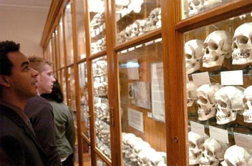
Most of all, this museum is known for its huge collection of skulls.
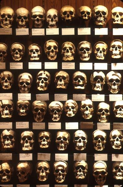
Among them there are unique exhibits, such as the human intestine length 12, 5 cm, and the corpse of a woman, who turned into a soap in the land where she was buried.
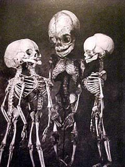
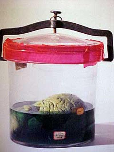
On display are a lot of wax models, as well as preserved organs and whole bodies ...
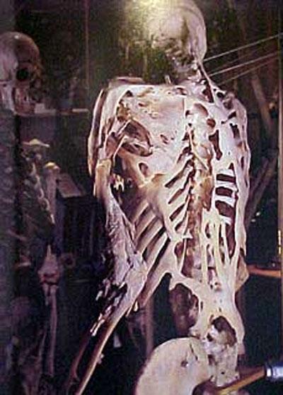
One of the main exhibits of the Museum Mutter - Garii Istlak, a man in his lifetime suffering ossificans progressive fibrodisplations (Fibrodysplasia Ossificans Progressiva, FOP) - a disease in which the formation of the extra bone in place of a bruise or a wound ...
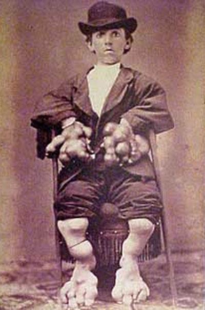
Before he died at the age of forty-odd years, Istlak donated to the museum by his skeleton ...
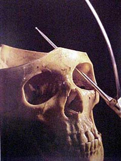
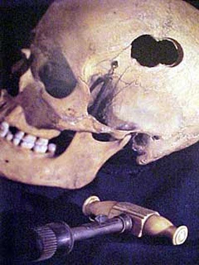
It is a tool for craniotomy. That such instruments in the previous century, a hole was drilled in the skull bone to remove blood clots from the cavity of the wound ...
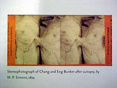
The famous Siamese twins Chang and Yong Bin (Cheng and Eng Bunker, 1811 - 1874) with a combined liver ...
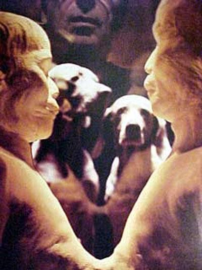
Photo of Siamese twins, made after the autopsy ...
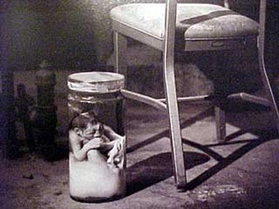
The skeleton-headed child ...
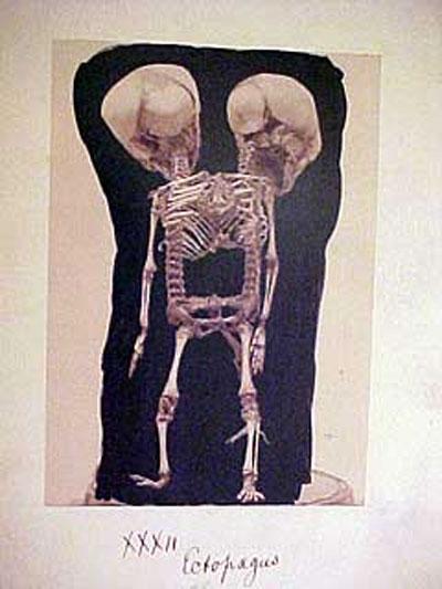
Skeleton of Siamese twins connected by their heads ...
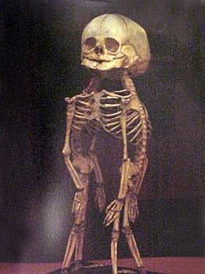
Mummified part of the pelvis to the lower limbs ...
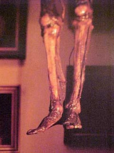
Mummified baby's head, with the manifestation of veins and arteries ...
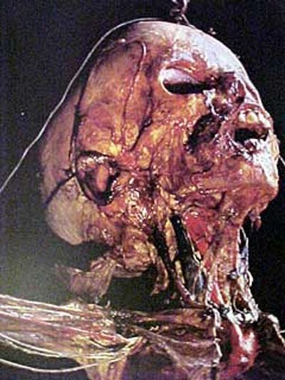
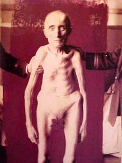
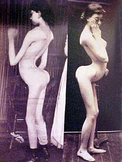
Woman with unusual curvature of the spine ... The development of the disease started in 2 years. For 20 years the disease has affected all the joints of the body, so that she could move only with the legs ...
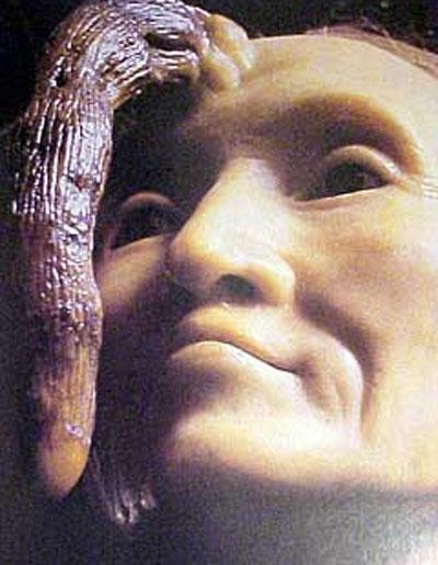
This woman - Madame Dimanche. She lived in Paris in the early nineteenth century. For 76 years on her forehead she grew horn and 82 years reaching a length of 20 cm. It has been successfully removed the famous French surgeon Joseph Suberbeillom (Joseph Souberbeille, 1754-1846) ...
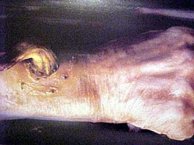
Hand with a similar growth on the wrist ...
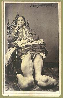
Pathology feet ...
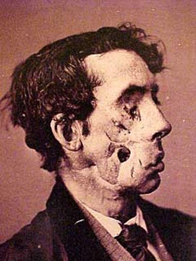
Harvey soldier was wounded in the Battle of Chancellorsville (Chancellorsville) May 3, 1863. A fragment of a projectile hit in the right eye ... So with the shot in the head, a small deafness and paralysis of the right side of privates lived until December 9, 1869. Died for unknown reasons ...
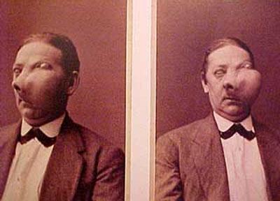
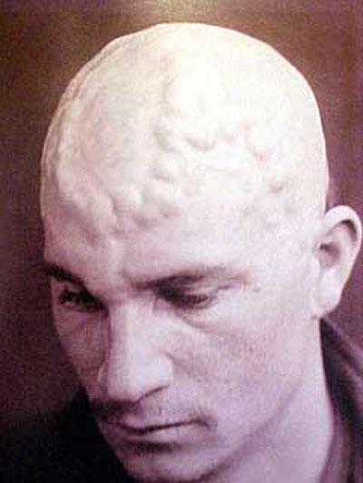
Unusual swelling on his head ...
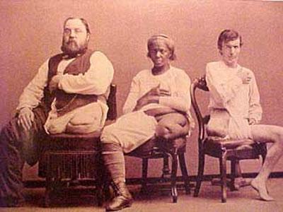
Successful cases of amputation of the feet ...
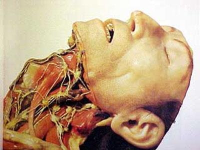
Dummy head with the facial nerve, and eye, modeled in wax, shows the relationship of the surface of facial nerve with the bones of the skull. Made in Paris around 1880 ...

Conjoined twins after opening ...
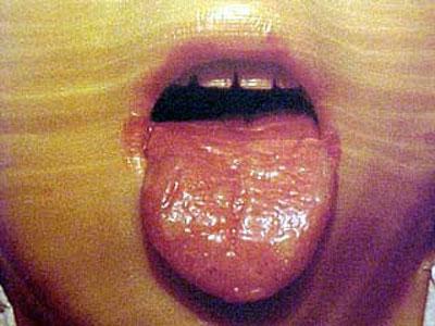
Syphilitic change the language ...
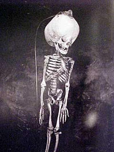
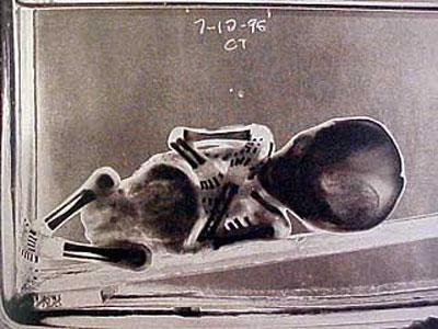
The baby with hydrocephalus skull ...
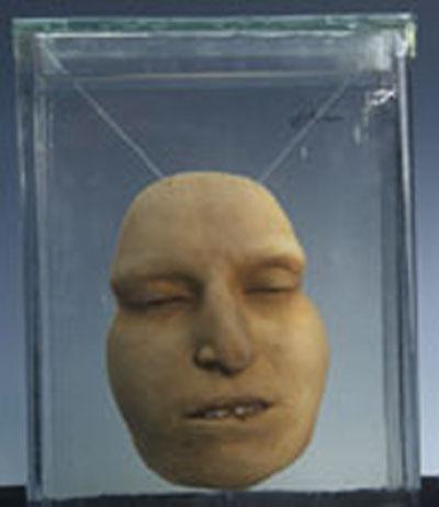
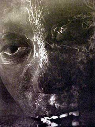
Face preserved with salicylic acid methyl and benzyl benzoate, which allow to make visible veins ...
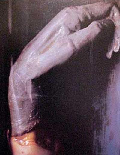
Dry gangrene hands ...
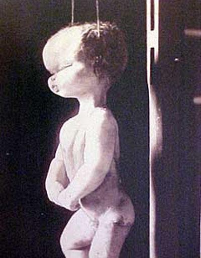
Twelve-fruit ...

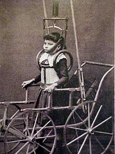
Paralysis of the legs ...
Cancer of the throat ...
Hydrocephalus skull ...
Trёhnogie Siamese twins ...
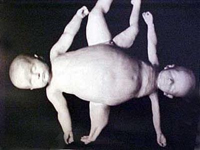
Fruit in glycerol ...
Varicose veins in the head ...
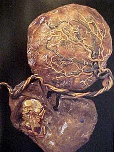
The placenta of twins ...
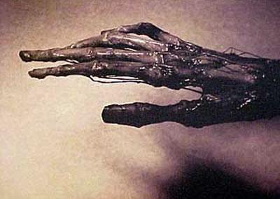
Dried hand ...
For centuries, medical practice improved ... The Mutter Museum for anyone interested in the history of medicine, there is something interesting ...
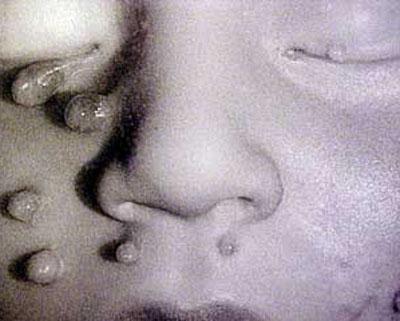
... Whether it's photos wounds of the Civil War the north and south, or combined liver famous Siamese twins Chang and Yong ...
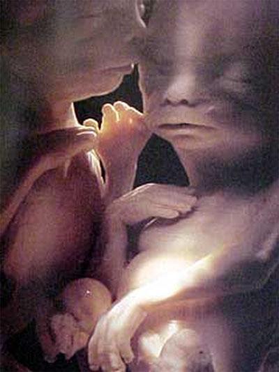
Look closely and ... pozaviduete themselves ... It's great that now the yard is not XVIII century!