2051
Lives of a Cell
Initially shows a blood vessel in which very quickly sweep erythrocytes, and slowed down his wall leukocyte.
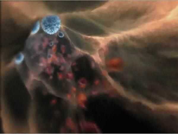
leukocyte "rolling" on the vascular endothelium ...
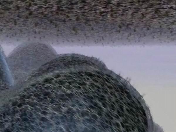
... And clings to different receptors for the corresponding receptors on the surface of endothelial cells.
There is a contact zone. This interaction will launch further events in the cell.
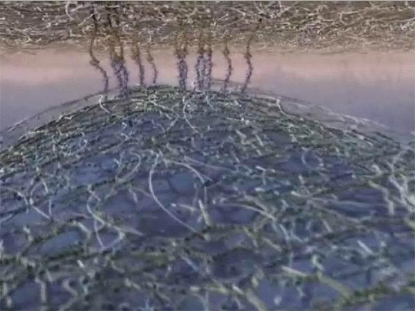
Next we see - already at a higher magnification - the cell membrane (apparently, all the same white blood cell). Membrane proteins involved in cell contact, focused on lipid rafts («lipid rafts»; I do not know, as they are now called in Russian, so that the ear does not hurt the - sorry).
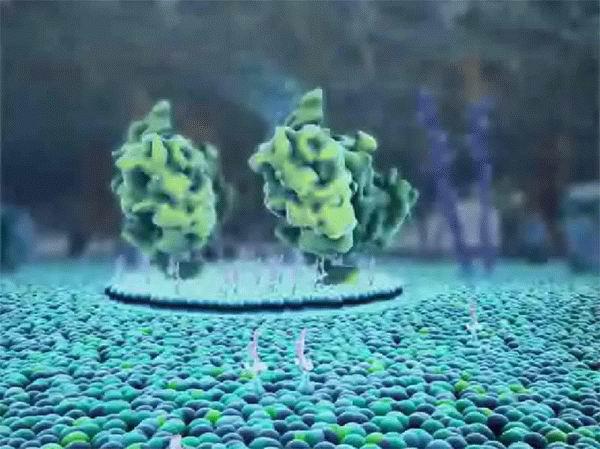
On the inner surface of the membrane, where the cells outside "snorting" receptors, signaling molecules are now collected. They will give a signal that the leukocyte came in contact with the endothelium, then - deep into the cells.
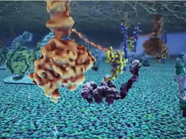
Similar to the membrane, inside view. If so, then this is probably the most endothelium than leukocyte.
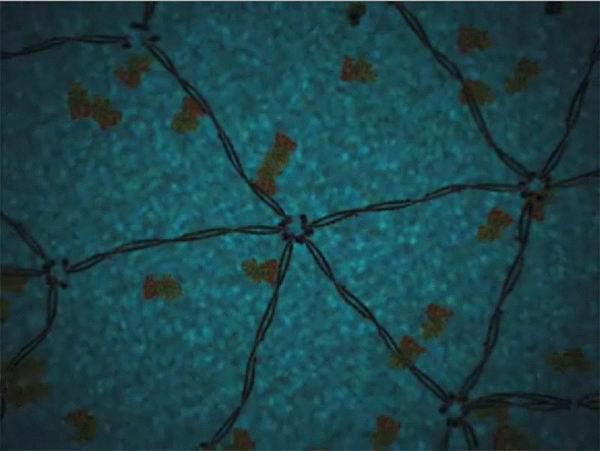
Showing different cytoskeletal elements forming the internal architecture of the cell.
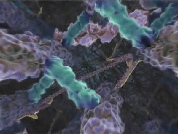
In the cell is going to actin filament.
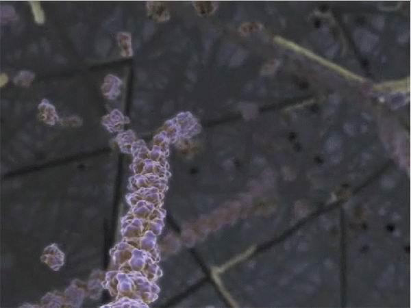
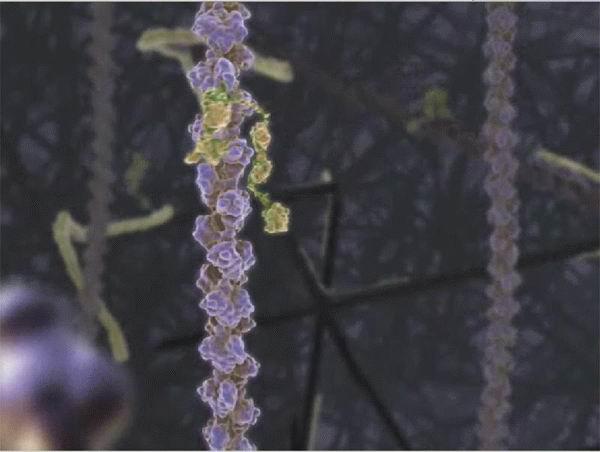
One of the actin-binding proteins actin microfilaments predatory bites.
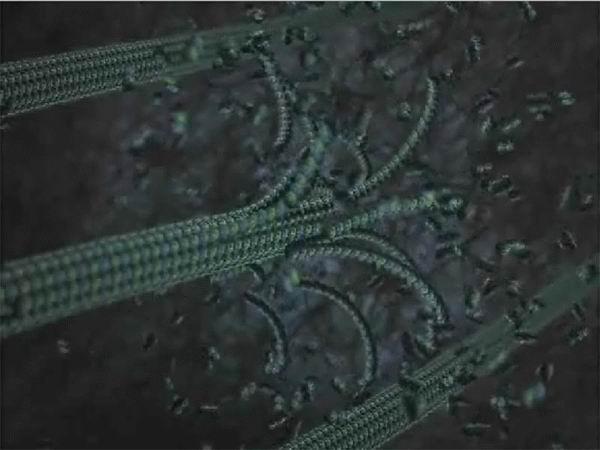
And here is going from tubulin microtubule ...
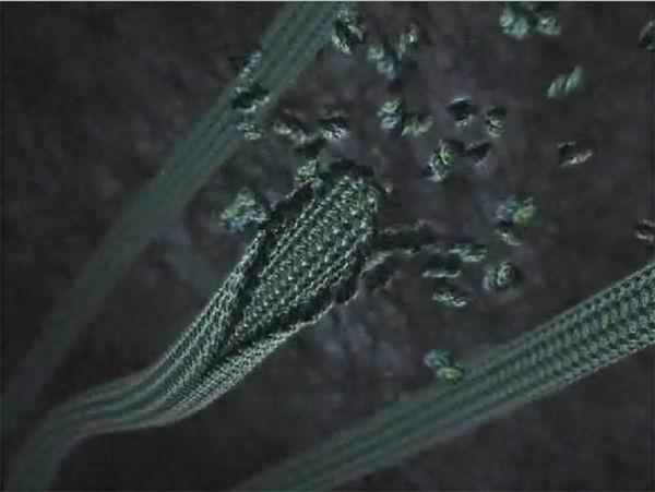
... And understands :) Microtubules are polar - going strictly on one end and the other dealt with.
One of the most memorable shots :) It is important to pace molecule that pulls a huge ball - apparently, kinesin (in dynein, IMHO, the "legs" would be larger and rounder, and he moved to the other side - in the following frames visible centriole) .
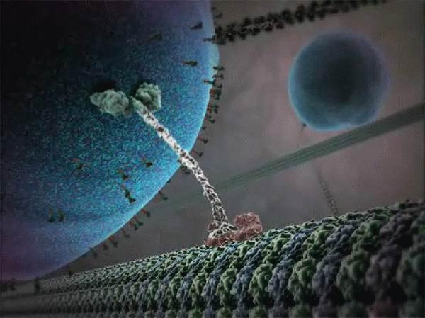
Kinesins move along microtubules in the cell and they are organized with the help of centrioles (the sphere inside which show two cylinder - this is what it is).
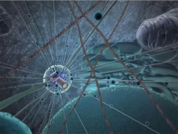
We are approaching the cell nucleus. Through pores in the nuclear envelope of the nucleus to the cytoplasm out of the matrix molecules (information) RNA.
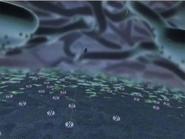
For mRNA in the cytoplasm join ribosomal subunit (shown in green) ...
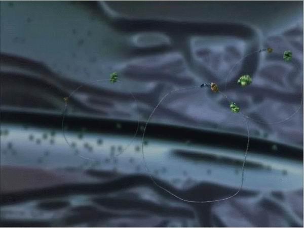
... And begins the synthesis of a protein molecule (meanders left), while the ribosome moves along the mRNA (long thread prodernutaya through the ribosome). When the synthesis is over, the protein particles of the ribosome and mRNA are separated.
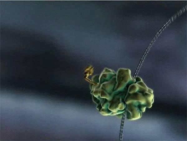
Shown here are two signaling molecules - hard to tell what was meant. Large gray shadow to which they float, similar to mitochondria, but not answer.
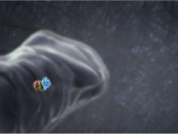
But a slightly different version of the events in the synthesis of protein. Ribosome and mRNA are on the surface of one of the endoplasmic reticulum vacuoles. Synthesized protein thus immediately gets into the vacuole rather than wandering through the cytoplasm.
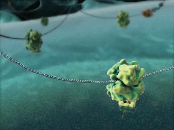
Pulsiruschie in the background round pieces - it seems vesicles of the endoplasmic reticulum. They are separated from the network and join the Golgi complex, carrying within themselves all sorts of things synthesized. And at the forefront again now
walking seem dynein / kinesin.
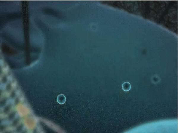
Here it is - the Golgi complex. This stack of membrane vesicles, one cleaves the bottom, while the other is separated from the top of the stack. While the contents of the bubble travels upward, it is subjected to various chemical modifications.
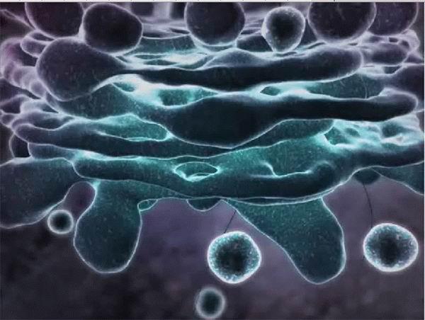
Separated from the Golgi complex vacuole inside dragged to the cell surface. Drags her all the same kinesin, it "from the back" is not visible, but we already know :)
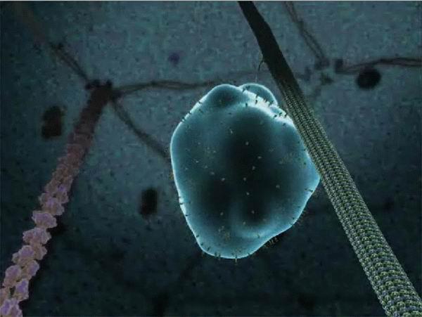
Merges vacuole membrane (not shown), and that was inside, spat out. Of the molecules are free to drift about their business, but some, it turns out to be secured inside the vacuole! Now they were attached to the outside of the cell membrane.
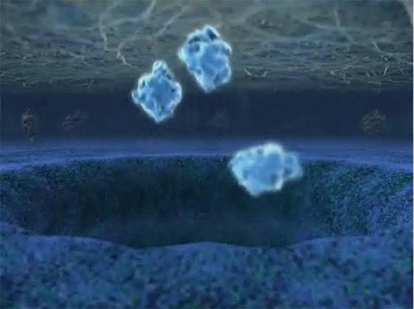
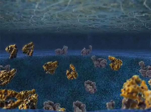
What hangs from the top - it's a different cell. She patiently waits for the same basal cell collects newly synthesized receptors on the same patch.
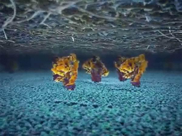
Under the snout meanwhile again formed already familiar lipid raft / raft.
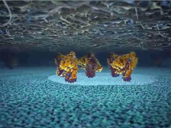
Adhesion molecules - receptors required for contact - activated "straightened," and the cells adhere tightly with each other.
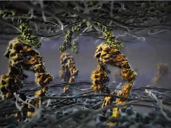
Leukocyte, kativshiysya on the endothelium (and rolling it, clinging to the receptors, as you already understood) finally stops and "flattens" to chose the place.
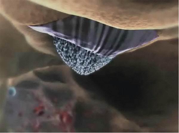
He squeezes between two endothelial cells, leaving the capillary. Endothelial cells at the same time have a little disconnected, but it is not shown.
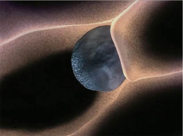
Actually, the Great planned by the director, apparently, was this - to show that in order to leukocyte could get out of the vessel in the tissue, it is necessary that on its surface, and on the surface of standing guard endothelial cell receptors present necessary, what can be nice dosintezirovat directly in the frame, simultaneously giving a general panoramic view different cell sites.
It was very nice, although the inaccuracies and exaggeration, of course, there are many. Undoubtedly, more or less reconciled the relative sizes of the interacting macromolecules. But now with reaction rates - seams that something is slow, something accelerated and kinesin, as all can see, just the same color before the camera :) But all the small rag-tag - ATP, amino acids, and other accessory proteins - from the frame driven, extra organelles draped, and instead of a thick pea soup to give a clear broth with single inclusions, eye-catching consumer :) For the sake of beauty and not a go ...
More images
- -