10 images that shocked the medical world
 Bashny.Net
On X-rays and tolko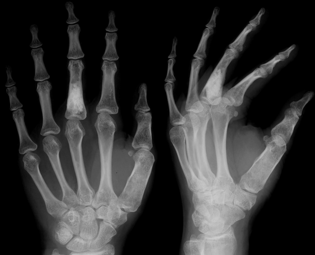
For most of us the passage of ultrasound, MRI, angiography, or X-ray is being in a windowless room, more like a prison than a hospital room. Specialist puts us in a special clothes, and then captures our body in various painful positions. At such moments, we are almost ready to see the torches on the walls, and the "iron maiden" in the corner. Here are 10 images that will make all of the above procedures are not so terrible.
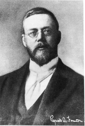
1. Engagement Ring Bertha Rentgen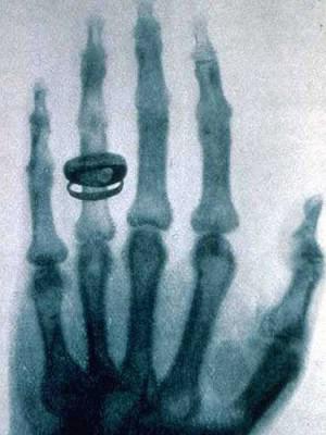
In November 1895, the physics professor Wilhelm Röntgen in Würzburg, Bavaria, studied the "electric rays" when he discovered that they can penetrate objects and project images of these objects on the fluorescent screen. When he put on the path of the rays his hand, I noticed that the picture showed the contrast between bone and translucent flesh. Roentgen immediately understood the consequences of his discovery: the doctors are now able to explore the human body without an autopsy. He replaced the fluorescent screen on a photographic plate and 8 November 1895 made the first X-ray. In the picture was a left hand of his wife, Bertha and her wedding ring.
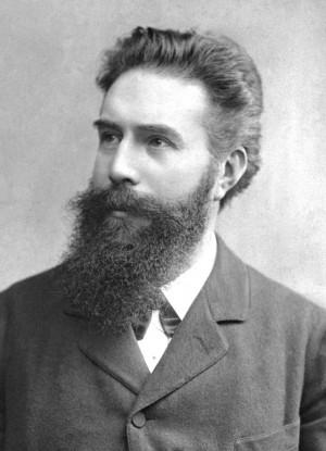
First, the world doubt about the opening of X-rays. In «The New York Times» Opening rejected, considering it a simple photographic technique known for a long time. But just a week later in «The Times» reports began to emerge of how X-rays are helpful to surgeons. One of these reports by a British doctor named John Hall-Edwards, who was the first to use X-rays to detect the needle hit the hand of the patient. In 1901 Röntgen was awarded the Nobel Prize in physics, and now its conclusions are considered to be "one of the greatest discoveries in the history of science».
2. Survey of the heart and digestive system by X luchey
After the discovery of X-rays the situation began to develop very quickly. Almost immediately, the researchers combined x-rays with film and began to record moving objects. The first in this matter was John McIntyre, a surgeon from the Royal Hospital in Glasgow. McIntyre has created the world's first X-ray department, and later it was in his office for the first time will find the stones in the kidney patient.
In 1897 McIntyre presented the Royal Society of London a short film. It was an X-ray frog legs. McIntyre took the foot, because in order to "educate" her, much less energy is required than for the "transmission" of a human foot. Later he took a beating human heart. In addition, he gave the patient bismuth and shot his digestive system to see how the absorption of bismuth.
Today, these films are called "X-ray". They are used to remove the placement of catheters in the heart, remove the digestive and urinary systems, and other medical procedures. In 2013, the year in the UK alone there were more than 1, 3 million fluoroscopic procedures.
3. Major Beevor hunts pulyami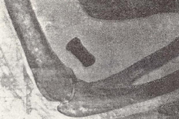
A few months after the discovery of X-rays have been used on the battlefield. They were first used during the Abyssinian War.
When Italy invaded Abyssinia in 1896, Lieutenant Colonel Giuseppe Alvaro used X-ray to look for a bullet in the forearm Italian soldiers. A year later, the X-rays were again used on the battlefield, but this time during the Greco-Turkish War. Despite many successes, the military did not hurry to recognize the effectiveness of the use of X-rays in the treatment of the wounded.
In June 1897, war broke out between Afghanistan and India. Britain sent its soldiers on the plateau Tirah to free mountain passes. Mayor Walter Beevor acquired x-ray equipment, and set it in a field hospital in the Tirah. He made more than 200 pictures, including a picture of the elbow of the Indian soldier with a bullet stuck there. Beevor also able to find the bullet lodged in his leg General Wodehouse. The following year, Beevor made a presentation before the scientific community, and since Britain became the full-time use on the battlefield X-ray machines. Other countries gradually began to follow the example of England.
One of the advantages of devices has been their portability. During the First World War, Marie Curie and her daughter Irene was brought to the front 20 x-ray machines in the trunk of a van. Today, mobile phones are used in the treatment of patients who are too ill to come to their own radiology department of the hospital.
4. Proof of damage to metal korsetami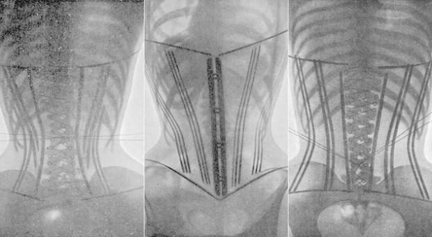
In one of the earliest known forms of medical imaging French physician Louis O'Follovel to draw attention to one issue, I made pictures of several women first torsos with metal corsets and then without them. The pictures clearly show how hard metal braces compress the chest, and there are internal organs. O'Follovel was not a supporter of a total ban corsets. He just wanted to be made more flexible. That is what happened in future. Pictures O'Follovela, supported the views of other influential physicians of the time, led to the fact that the industry began to produce more free corsets.
Later, however, the experts ask another question: Did the right O'Follovel use X-rays to prove a point? In those days, in order to take a picture, fixed object has been exposed beams for a long time. For example, to do in the 1896 picture of the forearm, it needed 45 minutes. To make the first dental x-ray film, it took 25 minutes. Women in corsets exposed beams twice as long, with most irradiated radiation-sensitive parts of the body: chest and abdomen (and hence the reproductive organs).
Danger radiation while already been well known. In the first year the test was recorded rays doctors hair loss, redness and peeling of the skin. Clarence Dally, working with rays for Thomas Edison, his hands repeatedly subjected to radiation, and it lasted for at least two years. Subsequently, both hands were amputated, and the Dali died of cancer in 1904. Most of the pioneers in the study of radiation (Mary and Irene Curie, John Hall-Edwards, Wilhelm Roentgen) died from diseases caused by radiation.
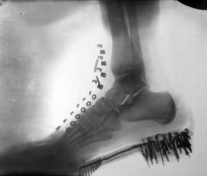
However, the world is not in a hurry to recognize the great danger of excess radiation. Women currently irradiated ovaries to treat depression. Radiation has been used as a remedy for ringworm, acne, impotence, arthritis, ulcer and even cancer. In beauty salons customers were exposed to did not grow facial hair. Irradiated toothpaste, chocolate, water. In 1920-1950-ies in many shoe stores were X-ray machines, taking pictures of feet in the shoes of customers to show how well she is sitting on her feet. Nowadays, X-rays are almost never used for non-medical purposes, however, and now is an increased risk of radiation. One study found that 18,500 cases of cancer worldwide medical x-ray is triggered.
5. The first kateter
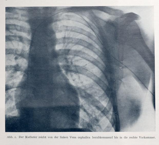
Surgeons at work in the hospital Victoria, Verner Forsman theorized that the flexible tube (catheter) can be inserted in the groin or arm through the veins of the patient and can be delivered directly into the heart bosom.
Most experts at the time decided that the catheter does not reach the heart in such a way, so the bosses in Victoria Hospital refused to give permission for this experiment doctor. Forsman But it did not scare, and he inserted a needle his left hand, and then the catheter is advanced along the main vein through the biceps, passing shoulder, directly into the heart. To do this, it took only 60 cm tube. Then Forsman down in X-ray department and took a picture to prove that the catheter really got to the heart. Later he performed the procedure on themselves several times. Unfortunately, Forsman made fun of his colleagues, considering the usual procedure of focus.
Discouraged, Forsman continued to work, retrain from a surgeon in urology. He did not know that the importance of his contribution to medicine will be recognized at once, so it was very puzzled when in October 1956 the year at his home phone rang, and he was informed that he had won the Nobel Prize in medicine and physiology. Dokotor simply asked: "Why»?
6. Giperfonografiya
One of the disadvantages of X-ray technology is that it allows us to see only images of dense anatomical structures, such as bone or foreign bodies (such as bullets). Another disadvantage is that radiation is dangerous, and it may well kill the child in the womb. So the medical world needed a secure way to display less dense structures of the body. The decision came after the collapse of the "Titanic" in 1912.
To better detect icebergs, Reginald Fessenden patented a device that emits sound waves directed and fixing their echoes reflected from various remote sites. His sonar was able to detect icebergs at a distance of two kilometers.
At the same time, the First World War broke out, and German submarines began to threaten Allied supply ships. The physicist Paul Langevin developed a hydrophone, which is also used sound waves to detect German submarines. April 23 1916, the year the boat was sunk by a German US-3. It was the first boat, detected by a hydrophone. After the war, the hydrophone technology used to detect defects in metals.
In the late 1930s, German psychiatrist and neurologist Karl Dussik believed that with the help of sound, you can look into the brain and look at other parts of the body that are not visible in X-rays. Dussik first to use sound for diagnostic purposes. Most of his work he has done in Austria. Later, he expanded and supplemented his studies, and then the world first heard the word "giperfonografiya».
And after ten years obstetrician from Scotland named Ian Donald borrowed an industrial ultrasound machine and used it to study a variety of tumors. Soon Donald started to successfully use this machine for the detection of malignant tumors and to monitor the status of the fetus in the womb.
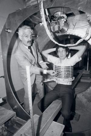
7. The first computer tomografiyaOdnim of the limitations of X-rays is that the picture appears all that is between the X-ray tube and the image itself. As a result, all kinds of pathologies such as tumors, can be hidden tissues, organs and bones located above or below.
In the 1930s, it began to flourish tomography. It was an X-ray of certain levels of the body, and all that is above or below the plane of the necessary, the picture looks blurred. This was done by moving the X-ray tube during the shooting. The tube can be moved in three planes of the human body: sagittal (left), coronal (front to back), and the center, she is cross-sectional plane (from the feet to the head).
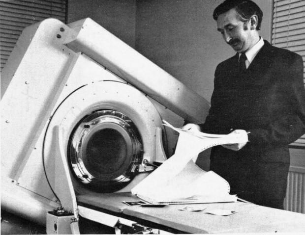
And in 1967, a scientist from the EMI named Godfrey Hounsfield invented axial tomography. EMI is also the record company, which has sold 200 million records group «The Beatles», so she used her money to finance the Hounsfield for four years. That's how much it took to create a prototype of the device. His films were used instead of the scanner sensor and the patient just passed between the tubes and sensors at a predetermined rate. After that the computer has revolutionized the anatomy of the patient. Today it is simply called: computerized tomography. October 1, 1971, the year used for the first time Hounsfield own invention for the detection of tumors in the brain of a woman.
8. The first magnetic resonance tomografiyaPri MRI machine creates a static magnetic field, which builds all the protons in the body in one direction. Then short bursts of radio waves displace these protons, and as soon as the radio waves are turned off, the computer measures the time it took to rebuild the protons. After that the computer uses these measurements to reconstruct the image of the patient's body.
It might appear that machines for computed tomography (CT) and magnetic resonance imaging (MRI) are very similar, but they are different. CT uses a potentially dangerous radiation, whereas MRI does not. In addition, MRI shows organs and soft tissue much better than CT. MRI is used when the doctor wants to see the condition of the spinal cord, ligaments and tendons. On the other hand, CT allows a better view of the spine and bone damage.
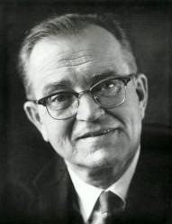
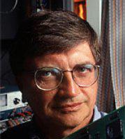
The first MRI scanner body came up with physicist Raymond Vahan Damadian in 1969. In 1971, he first published his theory about this device in the journal Science Magazine. In March 1972, Damadyan patented his invention. July 3, 1977, it was held the first MRI scan of man.
Since none of his colleagues did not want to go into a new scanner, Damadyan climbed there myself. When nothing worked, the staff suggested that their boss was too big. One of the participants graduate Larry Minkoff was slimmer and volunteered to try. In the picture above you can see a snapshot of breast Minkoff.
9. Laparoskopiya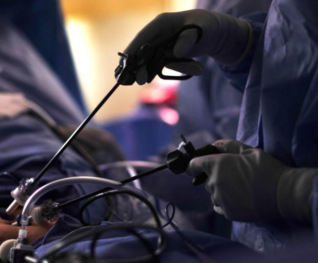
Surgeons throughout the centuries removed from the stomachs of people all sorts of things. And these bellies were always opened. This makes the patient very susceptible to infections, and for the recovery operation needed for a long time.
But in 1901 a gynecologist von Ott from Petrograd presented a laparoscopy - a method in which the operation is carried out not through the large hole, and through one or more small holes or cracks. The surgeon thus looking directly into the stomach or chest of the patient via the device, which in appearance resembles a miniature telescopes. Instead of having to use their hands, surgeons use forceps, scissors, clamps and other tools in a very long rods.
Unfortunately, this also means that the surgeon doing such operations must sometimes take the most unexpected postures, to see where it is needed. One surgeon once recalled that he had to lie down on the thigh of the patient to remove his gall bladder. And at 2, 5:00 this surgery, the doctor was completely exhausted. It is for this reason laparoscopy is of limited use.
10. three- and a four ultrasound
For thirty years, ultrasound has been limited to only two dimensions in which the devices first send sound waves, and then fixed echo. Millions of parents have tried valiantly, but were never able to make out in the black-and-white pictures, look how their child. Since 1970, scientists working on the three-dimensional ultrasonography (US) children. Sound waves are sent from different angles and in different directions, and then from the resulting echo reconstructed image of the child. Almost in the same way as does the scanner.
In 1984 Kazunori Baba from the Tokyo Institute of Medical Electronics was the first person who managed to get a three-dimensional image of a baby in the womb. But the image quality and the amount of time required for image reconstruction (10 minutes) made the diagnostic method is unsuitable.
In 1987, Olaf von Ramm and Steven Smith patented the first high-speed method of three-dimensional ultrasound, which will improve image quality and reduce the time of processing. And then the real boom of ultrasound, especially after the addition of four-dimensional ultrasound, in which parents are able to see the movement of their child.
via factroom.ru

For most of us the passage of ultrasound, MRI, angiography, or X-ray is being in a windowless room, more like a prison than a hospital room. Specialist puts us in a special clothes, and then captures our body in various painful positions. At such moments, we are almost ready to see the torches on the walls, and the "iron maiden" in the corner. Here are 10 images that will make all of the above procedures are not so terrible.
1. Engagement Ring Bertha Rentgen

In November 1895, the physics professor Wilhelm Röntgen in Würzburg, Bavaria, studied the "electric rays" when he discovered that they can penetrate objects and project images of these objects on the fluorescent screen. When he put on the path of the rays his hand, I noticed that the picture showed the contrast between bone and translucent flesh. Roentgen immediately understood the consequences of his discovery: the doctors are now able to explore the human body without an autopsy. He replaced the fluorescent screen on a photographic plate and 8 November 1895 made the first X-ray. In the picture was a left hand of his wife, Bertha and her wedding ring.

First, the world doubt about the opening of X-rays. In «The New York Times» Opening rejected, considering it a simple photographic technique known for a long time. But just a week later in «The Times» reports began to emerge of how X-rays are helpful to surgeons. One of these reports by a British doctor named John Hall-Edwards, who was the first to use X-rays to detect the needle hit the hand of the patient. In 1901 Röntgen was awarded the Nobel Prize in physics, and now its conclusions are considered to be "one of the greatest discoveries in the history of science».
2. Survey of the heart and digestive system by X luchey

After the discovery of X-rays the situation began to develop very quickly. Almost immediately, the researchers combined x-rays with film and began to record moving objects. The first in this matter was John McIntyre, a surgeon from the Royal Hospital in Glasgow. McIntyre has created the world's first X-ray department, and later it was in his office for the first time will find the stones in the kidney patient.
In 1897 McIntyre presented the Royal Society of London a short film. It was an X-ray frog legs. McIntyre took the foot, because in order to "educate" her, much less energy is required than for the "transmission" of a human foot. Later he took a beating human heart. In addition, he gave the patient bismuth and shot his digestive system to see how the absorption of bismuth.
Today, these films are called "X-ray". They are used to remove the placement of catheters in the heart, remove the digestive and urinary systems, and other medical procedures. In 2013, the year in the UK alone there were more than 1, 3 million fluoroscopic procedures.
3. Major Beevor hunts pulyami

A few months after the discovery of X-rays have been used on the battlefield. They were first used during the Abyssinian War.
When Italy invaded Abyssinia in 1896, Lieutenant Colonel Giuseppe Alvaro used X-ray to look for a bullet in the forearm Italian soldiers. A year later, the X-rays were again used on the battlefield, but this time during the Greco-Turkish War. Despite many successes, the military did not hurry to recognize the effectiveness of the use of X-rays in the treatment of the wounded.
In June 1897, war broke out between Afghanistan and India. Britain sent its soldiers on the plateau Tirah to free mountain passes. Mayor Walter Beevor acquired x-ray equipment, and set it in a field hospital in the Tirah. He made more than 200 pictures, including a picture of the elbow of the Indian soldier with a bullet stuck there. Beevor also able to find the bullet lodged in his leg General Wodehouse. The following year, Beevor made a presentation before the scientific community, and since Britain became the full-time use on the battlefield X-ray machines. Other countries gradually began to follow the example of England.

One of the advantages of devices has been their portability. During the First World War, Marie Curie and her daughter Irene was brought to the front 20 x-ray machines in the trunk of a van. Today, mobile phones are used in the treatment of patients who are too ill to come to their own radiology department of the hospital.
4. Proof of damage to metal korsetami

In one of the earliest known forms of medical imaging French physician Louis O'Follovel to draw attention to one issue, I made pictures of several women first torsos with metal corsets and then without them. The pictures clearly show how hard metal braces compress the chest, and there are internal organs. O'Follovel was not a supporter of a total ban corsets. He just wanted to be made more flexible. That is what happened in future. Pictures O'Follovela, supported the views of other influential physicians of the time, led to the fact that the industry began to produce more free corsets.
Later, however, the experts ask another question: Did the right O'Follovel use X-rays to prove a point? In those days, in order to take a picture, fixed object has been exposed beams for a long time. For example, to do in the 1896 picture of the forearm, it needed 45 minutes. To make the first dental x-ray film, it took 25 minutes. Women in corsets exposed beams twice as long, with most irradiated radiation-sensitive parts of the body: chest and abdomen (and hence the reproductive organs).
Danger radiation while already been well known. In the first year the test was recorded rays doctors hair loss, redness and peeling of the skin. Clarence Dally, working with rays for Thomas Edison, his hands repeatedly subjected to radiation, and it lasted for at least two years. Subsequently, both hands were amputated, and the Dali died of cancer in 1904. Most of the pioneers in the study of radiation (Mary and Irene Curie, John Hall-Edwards, Wilhelm Roentgen) died from diseases caused by radiation.

However, the world is not in a hurry to recognize the great danger of excess radiation. Women currently irradiated ovaries to treat depression. Radiation has been used as a remedy for ringworm, acne, impotence, arthritis, ulcer and even cancer. In beauty salons customers were exposed to did not grow facial hair. Irradiated toothpaste, chocolate, water. In 1920-1950-ies in many shoe stores were X-ray machines, taking pictures of feet in the shoes of customers to show how well she is sitting on her feet. Nowadays, X-rays are almost never used for non-medical purposes, however, and now is an increased risk of radiation. One study found that 18,500 cases of cancer worldwide medical x-ray is triggered.
5. The first kateter

Surgeons at work in the hospital Victoria, Verner Forsman theorized that the flexible tube (catheter) can be inserted in the groin or arm through the veins of the patient and can be delivered directly into the heart bosom.
Most experts at the time decided that the catheter does not reach the heart in such a way, so the bosses in Victoria Hospital refused to give permission for this experiment doctor. Forsman But it did not scare, and he inserted a needle his left hand, and then the catheter is advanced along the main vein through the biceps, passing shoulder, directly into the heart. To do this, it took only 60 cm tube. Then Forsman down in X-ray department and took a picture to prove that the catheter really got to the heart. Later he performed the procedure on themselves several times. Unfortunately, Forsman made fun of his colleagues, considering the usual procedure of focus.

Discouraged, Forsman continued to work, retrain from a surgeon in urology. He did not know that the importance of his contribution to medicine will be recognized at once, so it was very puzzled when in October 1956 the year at his home phone rang, and he was informed that he had won the Nobel Prize in medicine and physiology. Dokotor simply asked: "Why»?
6. Giperfonografiya

One of the disadvantages of X-ray technology is that it allows us to see only images of dense anatomical structures, such as bone or foreign bodies (such as bullets). Another disadvantage is that radiation is dangerous, and it may well kill the child in the womb. So the medical world needed a secure way to display less dense structures of the body. The decision came after the collapse of the "Titanic" in 1912.

To better detect icebergs, Reginald Fessenden patented a device that emits sound waves directed and fixing their echoes reflected from various remote sites. His sonar was able to detect icebergs at a distance of two kilometers.
At the same time, the First World War broke out, and German submarines began to threaten Allied supply ships. The physicist Paul Langevin developed a hydrophone, which is also used sound waves to detect German submarines. April 23 1916, the year the boat was sunk by a German US-3. It was the first boat, detected by a hydrophone. After the war, the hydrophone technology used to detect defects in metals.

In the late 1930s, German psychiatrist and neurologist Karl Dussik believed that with the help of sound, you can look into the brain and look at other parts of the body that are not visible in X-rays. Dussik first to use sound for diagnostic purposes. Most of his work he has done in Austria. Later, he expanded and supplemented his studies, and then the world first heard the word "giperfonografiya».
And after ten years obstetrician from Scotland named Ian Donald borrowed an industrial ultrasound machine and used it to study a variety of tumors. Soon Donald started to successfully use this machine for the detection of malignant tumors and to monitor the status of the fetus in the womb.
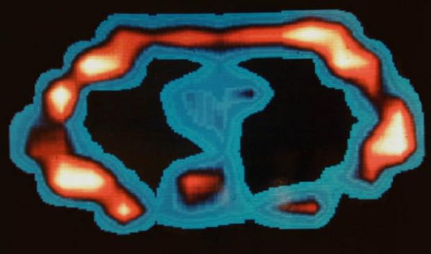
7. The first computer tomografiyaOdnim of the limitations of X-rays is that the picture appears all that is between the X-ray tube and the image itself. As a result, all kinds of pathologies such as tumors, can be hidden tissues, organs and bones located above or below.
In the 1930s, it began to flourish tomography. It was an X-ray of certain levels of the body, and all that is above or below the plane of the necessary, the picture looks blurred. This was done by moving the X-ray tube during the shooting. The tube can be moved in three planes of the human body: sagittal (left), coronal (front to back), and the center, she is cross-sectional plane (from the feet to the head).

And in 1967, a scientist from the EMI named Godfrey Hounsfield invented axial tomography. EMI is also the record company, which has sold 200 million records group «The Beatles», so she used her money to finance the Hounsfield for four years. That's how much it took to create a prototype of the device. His films were used instead of the scanner sensor and the patient just passed between the tubes and sensors at a predetermined rate. After that the computer has revolutionized the anatomy of the patient. Today it is simply called: computerized tomography. October 1, 1971, the year used for the first time Hounsfield own invention for the detection of tumors in the brain of a woman.
8. The first magnetic resonance tomografiyaPri MRI machine creates a static magnetic field, which builds all the protons in the body in one direction. Then short bursts of radio waves displace these protons, and as soon as the radio waves are turned off, the computer measures the time it took to rebuild the protons. After that the computer uses these measurements to reconstruct the image of the patient's body.

It might appear that machines for computed tomography (CT) and magnetic resonance imaging (MRI) are very similar, but they are different. CT uses a potentially dangerous radiation, whereas MRI does not. In addition, MRI shows organs and soft tissue much better than CT. MRI is used when the doctor wants to see the condition of the spinal cord, ligaments and tendons. On the other hand, CT allows a better view of the spine and bone damage.
The first MRI scanner body came up with physicist Raymond Vahan Damadian in 1969. In 1971, he first published his theory about this device in the journal Science Magazine. In March 1972, Damadyan patented his invention. July 3, 1977, it was held the first MRI scan of man.

Since none of his colleagues did not want to go into a new scanner, Damadyan climbed there myself. When nothing worked, the staff suggested that their boss was too big. One of the participants graduate Larry Minkoff was slimmer and volunteered to try. In the picture above you can see a snapshot of breast Minkoff.
9. Laparoskopiya

Surgeons throughout the centuries removed from the stomachs of people all sorts of things. And these bellies were always opened. This makes the patient very susceptible to infections, and for the recovery operation needed for a long time.
But in 1901 a gynecologist von Ott from Petrograd presented a laparoscopy - a method in which the operation is carried out not through the large hole, and through one or more small holes or cracks. The surgeon thus looking directly into the stomach or chest of the patient via the device, which in appearance resembles a miniature telescopes. Instead of having to use their hands, surgeons use forceps, scissors, clamps and other tools in a very long rods.
Unfortunately, this also means that the surgeon doing such operations must sometimes take the most unexpected postures, to see where it is needed. One surgeon once recalled that he had to lie down on the thigh of the patient to remove his gall bladder. And at 2, 5:00 this surgery, the doctor was completely exhausted. It is for this reason laparoscopy is of limited use.
10. three- and a four ultrasound
For thirty years, ultrasound has been limited to only two dimensions in which the devices first send sound waves, and then fixed echo. Millions of parents have tried valiantly, but were never able to make out in the black-and-white pictures, look how their child. Since 1970, scientists working on the three-dimensional ultrasonography (US) children. Sound waves are sent from different angles and in different directions, and then from the resulting echo reconstructed image of the child. Almost in the same way as does the scanner.
In 1984 Kazunori Baba from the Tokyo Institute of Medical Electronics was the first person who managed to get a three-dimensional image of a baby in the womb. But the image quality and the amount of time required for image reconstruction (10 minutes) made the diagnostic method is unsuitable.

In 1987, Olaf von Ramm and Steven Smith patented the first high-speed method of three-dimensional ultrasound, which will improve image quality and reduce the time of processing. And then the real boom of ultrasound, especially after the addition of four-dimensional ultrasound, in which parents are able to see the movement of their child.
via factroom.ru
Tags
See also
A selection of iconic images that shocked the world
Amazing dog photos that touched the whole world
20 fotofalsifikatsy that spread all over the world
25 fearless women who forever changed the course of history. You say the weaker sex?
10 pseudo-discoveries that shocked the scientific world
Technical and scientific myths. Why planes fly
When you change the world, and no one noticed
Dave Gahan: man, crying in which half of the world ...
Strange and crazy Easter traditions in the world
25 indescribably beautiful minerals and rocks, which is reflected in the whole world.