800
Science Beauty (12 photos)
The American Society of Advancement of Science (AAAS), among other of its activities, publishes the journal Science, announced the results of the sixth annual competition of scientific photographs, illustrations, posters, cartoons and interactive educational projects.
"The Glass Forest»
Mario Stephanie (Second University of Naples, Italy)
1st place in the competition photos
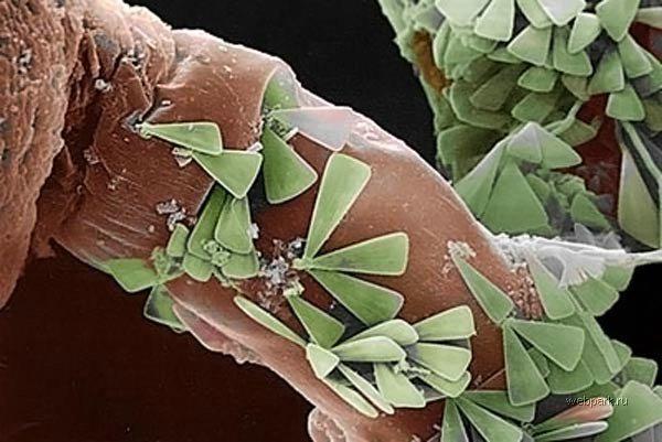
Diatoms - tiny protozoa which nevertheless produce about 40% of the oxygen in the world. Cells of these amazing beings enclosed in microscopic "shell" of the amorphous silica SiO2, which is whether the size of the sink are not 30 microns, as depicted in this photograph individuals Licmophora ehrenbergii, a few centimeters, we would call the glass.
Total science knows about 100,000 species of diatoms, the size of the largest of which reach 2 mm. In this photo, taken by a scanning electron microscope, shown in green diatoms strengthened on the "legs" of a small marine invertebrate Eudendrium racemosum, represented by shades of brown (the color in his usual sense of the electron microscope does not feel, as does not work with light, and with the flow electrons).
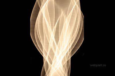
"String vibration»
Andrew Devidheyzi (Rochester Institute of Technology, USA)
2-4 places in the competition photos
As acknowledged by the photographer, he was going to capture the movement of winding elastic cotton thread, but overdid it with the speed of rotation of the motor, drive it into motion. As a result, the thread excited unusual rotational oscillations, the dynamics of which was much more difficult and picturesque than you can see, untwisted hanging chain. Picture was taken by a digital camera Canon delayed for about 2 seconds; During this time, the thread again managed to crank 10-20.
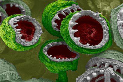
"Suckers squid: Little Monsters feed the beast»
Jessica Schiffman and Caroline Shouer (named Anthony Drexel University, USA)
2-4 places in the competition photos.
Tiny white "fangs" of chitin is at the end of suction cups that cover the "palm" of hunting long tentacles of squid. It is their squid prey capture and send it in her mouth. Diameter depicted in the electron micrograph of this sucker is about 0 to 4 mm, but most large squid may reach 5 cm. Color, recognized authors, they blew Frank Oz film "Little Shop of Horrors».
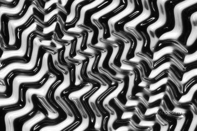
"The polymeric labyrinth»
Ye Eun Jin and Ueybel Douglas (University of Wisconsin in Madison, USA)
2-4 places in the competition photos.
Mixing polyethylene glycol and polidimetilsiloksan, Korean graduate student Ye Jin Eun, working under the biochemist Douglas Ueybela, was, as she puts it, "a polymer sandwich." The magic begins when the water gets sandwich. Polyethylene glycol while expanding, and its more hard polydimethylsiloxane copolymer - no, so that the first has no choice as to protrude upward, forming ridges and valleys, which are captured Eun using a digital camera and Nicon stereoscope Zeiss. By the way, was originally intended to secure the sandwich on it cell cultures. The experiment did not come out: such a wrinkled substrate nobody needs - but very beautiful.
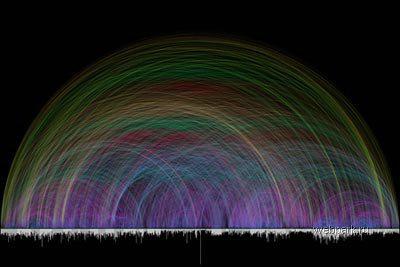
"Biblical visualization»
Chris Harrison (University named after Andrew Carnegie and Andrew Mellon and Richard, USA) and Christoph Römhild (Evangelical Lutheran Church of North Elba, Germany)
2-3 places in the competition of illustrations
This figure - the result of work of the German Christoph Römhild, make a complete (in his opinion) catalog of references from one chapter of the Bible (both Old Testament and New) on the other. In the submission of the form is the abundance of data transferred a computer program written by Harrison. Each chapter of each book of the Bible is represented by a column whose length reflects the number of verses in this chapter. Each arc - the same link to a catalog which have Römhild left more than one year; At the same time, the figure shows only those links that lead from one book to another - vnutriknizhnyh links from one chapter to another is not here.
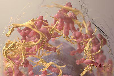
"Three-dimensional image of mammalian cells by scanning electron microscopy with ion stripping»
Donald Bliss and Sriram Subramanian (National Library of Medicine, National Institutes of Health, USA)
2-3 places in the competition of illustrations
This "dance" - in fact, represents the nucleus of the cancer cell. Her three-dimensional model obtained with an electron microscope, which is layer upon layer of scanned items, naked short bombardment of gallium ions, each time undermines the film thickness of 20 nm. This image shows the mitochondria (pink) and endoplasmic grid cell (gold) surrounding the core of black melanoma cells.
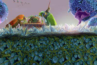
& Quot; «Mad Hatter tea party in" from Alice in Wonderland microscopic & quot;
Colleen and Dennis Kunkel Chemp (Concise Image Studios, USA)
1st place in the competition infographic
This illustration is for the journey to Alice in Wonderland, happen it to become even smaller than it occurred to Lewis Carroll - not a photograph. But it is made from photographs taken with different microscopes Dennis Kunkel. Colleen Chemp paint them and folded them from the whole picture. The painting beetles sit at the table of a butterfly's wing, which stands on the grass of the crystals of vitamin C, the roots of which mold filaments way through the same mold spores; mold here even the trees and the sky flying tiny aphids. Scale is not respected.
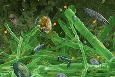
"Microbiology stream: Life in the biofilm»
Dofiyd Andrew and Gillian Lewis (University of Auckland, New Zealand)
2nd place in the competition infographic
A thin layer of green or brown mucus covering the stones in streams - complex ecosystems, the study of which in mountain streams of the North Island of New Zealand Andrew Dofiyd engaged under the direction of Gillian Lewis. People to better understand the object of his research, Dofiyd created this masterpiece of infographics with different magnification showing all the main inhabitants of the biofilm. This algae and fungi, bacteria and protozoa, blue-green algae (cyanobacteria) and even viruses. All of them accompanied by a short explanation of the role of these living (well, except for viruses sure) things in life biofilms. By the way, the poster is already in use in New Zealand schools.
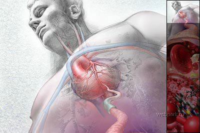
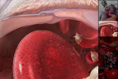
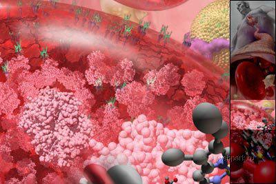
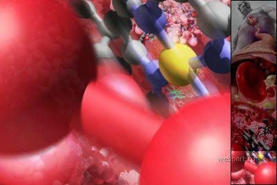
"Looking into the artery»
Linda Nye (Laboratory visualization museum The Exploratorium in San Francisco, USA)
1st place in the competition of illustrations
Little prospect of non-standard human circulatory system shows on the background of the heart and arteries of the human body of its blood cells, filled with hemoglobin, and even individual oxygen atoms in the structure of the fortified circulatory molecule. At each level of the image, in which a man was generally somewhere near the horizon, the magnification increases tenfold.
nauka_krasoty
"The Glass Forest»
Mario Stephanie (Second University of Naples, Italy)
1st place in the competition photos

Diatoms - tiny protozoa which nevertheless produce about 40% of the oxygen in the world. Cells of these amazing beings enclosed in microscopic "shell" of the amorphous silica SiO2, which is whether the size of the sink are not 30 microns, as depicted in this photograph individuals Licmophora ehrenbergii, a few centimeters, we would call the glass.
Total science knows about 100,000 species of diatoms, the size of the largest of which reach 2 mm. In this photo, taken by a scanning electron microscope, shown in green diatoms strengthened on the "legs" of a small marine invertebrate Eudendrium racemosum, represented by shades of brown (the color in his usual sense of the electron microscope does not feel, as does not work with light, and with the flow electrons).

"String vibration»
Andrew Devidheyzi (Rochester Institute of Technology, USA)
2-4 places in the competition photos
As acknowledged by the photographer, he was going to capture the movement of winding elastic cotton thread, but overdid it with the speed of rotation of the motor, drive it into motion. As a result, the thread excited unusual rotational oscillations, the dynamics of which was much more difficult and picturesque than you can see, untwisted hanging chain. Picture was taken by a digital camera Canon delayed for about 2 seconds; During this time, the thread again managed to crank 10-20.

"Suckers squid: Little Monsters feed the beast»
Jessica Schiffman and Caroline Shouer (named Anthony Drexel University, USA)
2-4 places in the competition photos.
Tiny white "fangs" of chitin is at the end of suction cups that cover the "palm" of hunting long tentacles of squid. It is their squid prey capture and send it in her mouth. Diameter depicted in the electron micrograph of this sucker is about 0 to 4 mm, but most large squid may reach 5 cm. Color, recognized authors, they blew Frank Oz film "Little Shop of Horrors».

"The polymeric labyrinth»
Ye Eun Jin and Ueybel Douglas (University of Wisconsin in Madison, USA)
2-4 places in the competition photos.
Mixing polyethylene glycol and polidimetilsiloksan, Korean graduate student Ye Jin Eun, working under the biochemist Douglas Ueybela, was, as she puts it, "a polymer sandwich." The magic begins when the water gets sandwich. Polyethylene glycol while expanding, and its more hard polydimethylsiloxane copolymer - no, so that the first has no choice as to protrude upward, forming ridges and valleys, which are captured Eun using a digital camera and Nicon stereoscope Zeiss. By the way, was originally intended to secure the sandwich on it cell cultures. The experiment did not come out: such a wrinkled substrate nobody needs - but very beautiful.

"Biblical visualization»
Chris Harrison (University named after Andrew Carnegie and Andrew Mellon and Richard, USA) and Christoph Römhild (Evangelical Lutheran Church of North Elba, Germany)
2-3 places in the competition of illustrations
This figure - the result of work of the German Christoph Römhild, make a complete (in his opinion) catalog of references from one chapter of the Bible (both Old Testament and New) on the other. In the submission of the form is the abundance of data transferred a computer program written by Harrison. Each chapter of each book of the Bible is represented by a column whose length reflects the number of verses in this chapter. Each arc - the same link to a catalog which have Römhild left more than one year; At the same time, the figure shows only those links that lead from one book to another - vnutriknizhnyh links from one chapter to another is not here.

"Three-dimensional image of mammalian cells by scanning electron microscopy with ion stripping»
Donald Bliss and Sriram Subramanian (National Library of Medicine, National Institutes of Health, USA)
2-3 places in the competition of illustrations
This "dance" - in fact, represents the nucleus of the cancer cell. Her three-dimensional model obtained with an electron microscope, which is layer upon layer of scanned items, naked short bombardment of gallium ions, each time undermines the film thickness of 20 nm. This image shows the mitochondria (pink) and endoplasmic grid cell (gold) surrounding the core of black melanoma cells.

& Quot; «Mad Hatter tea party in" from Alice in Wonderland microscopic & quot;
Colleen and Dennis Kunkel Chemp (Concise Image Studios, USA)
1st place in the competition infographic
This illustration is for the journey to Alice in Wonderland, happen it to become even smaller than it occurred to Lewis Carroll - not a photograph. But it is made from photographs taken with different microscopes Dennis Kunkel. Colleen Chemp paint them and folded them from the whole picture. The painting beetles sit at the table of a butterfly's wing, which stands on the grass of the crystals of vitamin C, the roots of which mold filaments way through the same mold spores; mold here even the trees and the sky flying tiny aphids. Scale is not respected.

"Microbiology stream: Life in the biofilm»
Dofiyd Andrew and Gillian Lewis (University of Auckland, New Zealand)
2nd place in the competition infographic
A thin layer of green or brown mucus covering the stones in streams - complex ecosystems, the study of which in mountain streams of the North Island of New Zealand Andrew Dofiyd engaged under the direction of Gillian Lewis. People to better understand the object of his research, Dofiyd created this masterpiece of infographics with different magnification showing all the main inhabitants of the biofilm. This algae and fungi, bacteria and protozoa, blue-green algae (cyanobacteria) and even viruses. All of them accompanied by a short explanation of the role of these living (well, except for viruses sure) things in life biofilms. By the way, the poster is already in use in New Zealand schools.

"Looking into the artery»
Linda Nye (Laboratory visualization museum The Exploratorium in San Francisco, USA)
1st place in the competition of illustrations
Little prospect of non-standard human circulatory system shows on the background of the heart and arteries of the human body of its blood cells, filled with hemoglobin, and even individual oxygen atoms in the structure of the fortified circulatory molecule. At each level of the image, in which a man was generally somewhere near the horizon, the magnification increases tenfold.


nauka_krasoty