Published the most detailed images of nerves
 Bashny.Net
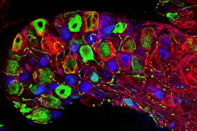
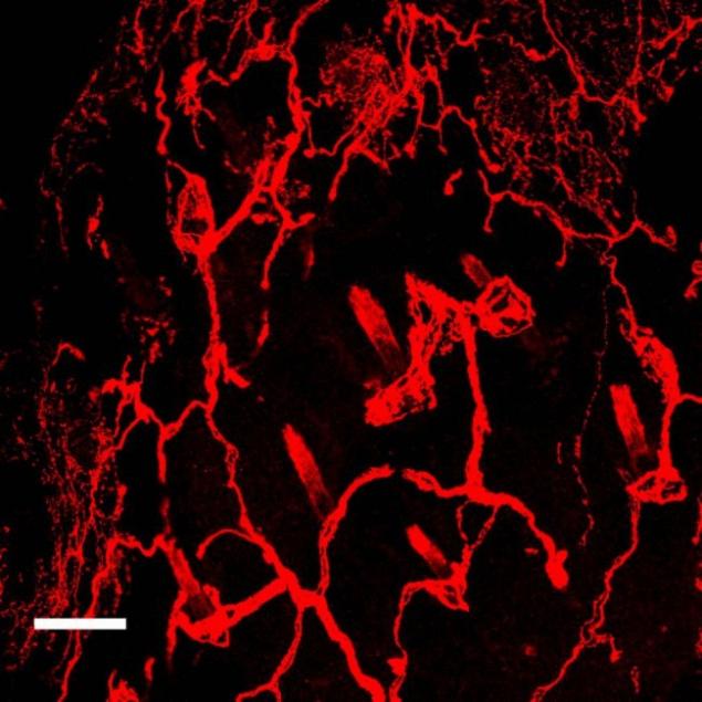
Scientists have published the most detailed pictures nerve endings mice, first used to living neurons technique a decade ago. The images show individual nerves, touch receptors and hair follicles, painted in bright fluorescent colors. All this is more like a modern abstract art, rather than on advanced microscopy.
Now you can see what we've seen before. High resolution allows to consider the individual elements in the background, reported the Verge Heppenstall Floor of the European molecular biology laboratory (EMBL) in Italy.
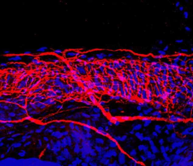
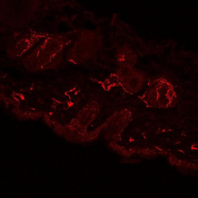
Heppenstall said that his favorite photo is one that shows hundreds of nerve endings located directly under the skin (the top-most image below).
Image obtained by the experts of the European molecular biology laboratory, impressive though that, despite the simplicity of collection and storage in the laboratory, skin samples are resistant to analysis by microscopy.
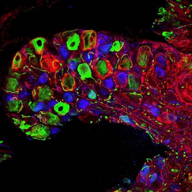
The problem is that it is extremely impermeable fabric. It is very difficult to introduce antibodies and conventional markers. It also has background fluorescence. Therefore, if you illuminate tissue with blue light in normal imaging], it fluoresces green. In the result, it is impossible to see anything — explained the researcher.
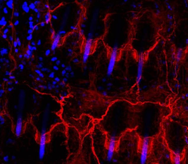
The technique of the so-called SNAP-tagging solves this problem by using a special protein, forming an artificial form that can pass through the skin barrier and be visible on a fluorescent background. This combination of chemical and biological properties, we can observe stunning images.
The work of scientists does not end there. Heppenstall said that his ultimate goal is to detect individual neurons and to capture them in action.
I'd like to look in the brain or in the spinal cord. First, to lighten the fabric, and then see neural circuits. Now we just observe that they are. But we also want to see how the nerve fibers are transmitted signals.
Source: hi-news.ru
Tags
See also
10 most interesting images of the microscopic world («Science-Photo»)
Published new pictures of Pluto the satellites of NYX and Hydra
Creepy portraits of women
Live photos
Wonders of Photoshop
Live photos
20 images, which look says more than any words
Hubble made detailed images of Pluto
Dawn continues to explore white spots Ceres