Triggers: Map of points of pain and tension in the body
 Bashny.Net
Designations in the figures:
Solid red shows the basic area of pain, granular - additional zone.
Crosses marked trigger points (stress points).
Head and neck
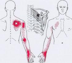
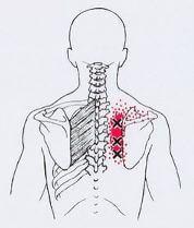
Trapezoidal muscle
Pattern of referred pain and localization of trigger points in the upper part of trapeze muscle.
Sternocleidomastoid muscle
Pattern of referred pain and localization of the responsible trigger points in the right sternocleidomastoid muscle. On the left of the sternum (surface) portion. Right — clavicular (deep) portion.
The masseter
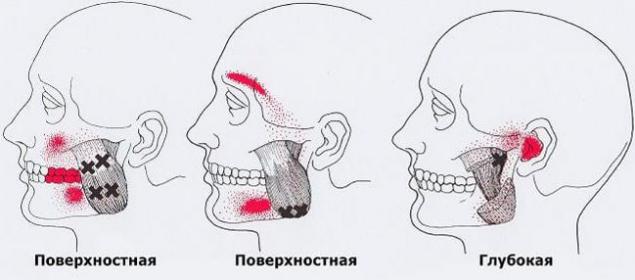
Localization of trigger points in various parts of the chewing muscles. Solid red shows the basic area of pain, granular — additional zone. Left — surface layer, the upper and middle division. In the center — superficial layer, lower part. Right — deep layer, upper part, just below the temporomandibular joint.
The temporalis muscle
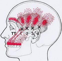
The picture of the reflected pain from TRIG-guérin dots in the left temporal muscle. Solid red shows the basic area of pain, granular — additional zone. Front "needle" pain arises from the anterior fibers (ТТ1), the middle "spokes" of ТТ2 and TTZ, rear (nadolna) "spoke" of ТТ4.
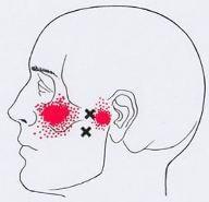
Medial hyoid muscle
Picture otrajenii pain (marked in red) and localization of the responsible trigger points in the medial hyoid muscle. Left — the outer area of pain, which can indicate patients. Right — the picture of the internal part of the pain was passing through the temporomandibular joint.
The lateral hyoid muscle
Pattern of referred pain from trigger points in the lateral hyoid muscle.
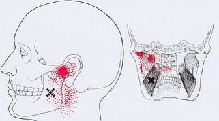
Digastric
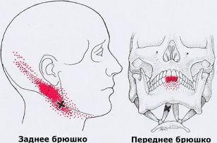
Picture of trigger points, and reflected from them, pain in the right dobrydney muscle.
Left — back abdomen — side view. Right — front abdomen — front view.
Suboccipital muscle
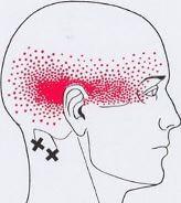
The picture reflected pain and trigger points in the right suboccipital muscle.
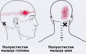
Pattern of referred pain (marked in red) and location of trigger points in the muscles polyostotic. Left upper trigger point in the muscle polyostotic head. To the right is the trigger point in the third layer polyostotic muscles of the neck.
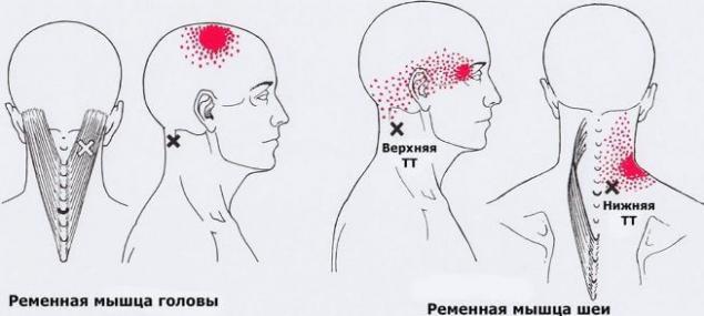
Trigger points and pattern they cause pain in the right belt muscle of the head and neck. On the left drawings — trigger points in the belt muscles of the head, in the occipital triangle. The figures on the right — upper trigger point that causes pain in the eye orbits, the lower the trigger point that causes pain in the angle of the neck.
Shoulders, chest and arms
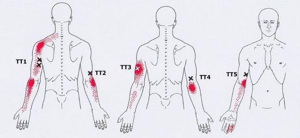
Trapezoidal muscle
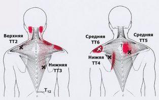
Referred pain and localization ТТ2 in the upper part of trapeze muscle, TTZ, ТТ4 at the bottom, ТТ5, TTB — in secondary departments trapezoidal muscles.
Muscle, levator scapulae
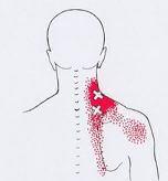
Picture of mixed pain, caused by two trigger points in the right muscle, levator scapulae. Solid red shows the basic area of pain, granular — additional zone.
Scalene muscle
A complex picture of pain caused by trigger points located in the anterior, middle and posterior scalene muscles. Some trigger points may have only one ongoing area otrajenii pain.
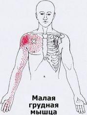
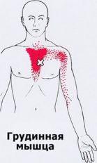
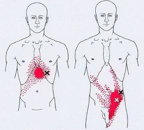
The pectoralis major muscle
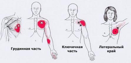
Pattern of referred pain and localization of trigger points in the pectoralis major muscle.
On the leftmost figure shows how overlapped the pain referred from the two trigger points located in the middle part of the muscle around of the sternum. Further, the figures show: left — the trigger point in the intermediate part Grudin-tion division, in the center — of TT in the clavicular part to the right is the trigger point of the free edges of the muscle that forms the axilla.
Trigger points in the right pectoralis minor muscle and its pain pattern caused.
Pattern of referred pain caused by trigger point in the left breast muscle.
Subclavian muscle
Pattern of referred pain caused by trigger point in the right subclavian muscle.
The serratus anterior muscle
Pattern of referred pain caused by trigger point localized in the right serratus anterior. Side view, back and front.
Rear-upper serrated muscle
Pattern of referred pain from trigger points in the right-rear-upper serrated muscle. Areas of persistent pain is marked by a solid red. Granular marked areas of possible pain. At left — back. The figure in the center of the blade assigned forward and the trigger point is available for palpation and injection. On the right picture the front view.
Rear-lower serrated muscle
Pattern of referred pain from trigger points in the right-rear-lower serrated muscle.
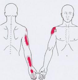
Latissimus dorsi
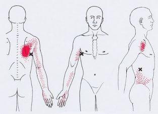
Pattern of referred pain and trigger points in the right-the widest muscle in the back. To the left is the usual localization of trigger points in the axillary portions of the muscle. In the center — front view. Right — the picture of pain from the lower trigger point.
Supraspinatus muscle
Pattern of referred pain and localization of trigger points in the right supraspinatus muscle.
Infraspinatus muscle
The picture reflected the pain and localization of trigger points in the right infraspinatus muscle.
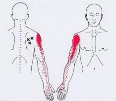
Small round muscle
Pattern of referred pain and localization of trigger points in the right of the small round muscle.
Large round muscle
Medial and lateral (back and axillary) trigger points in the right a large circular muscle and the pattern of reflected pain. Left — medial trigger points, the right — lateral TT.
Subscapularis muscle
Pattern of referred pain from trigger points in the right subscapularis muscle.
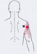
Rhomboid muscle
The overall picture of pain from trigger points in the right rhomboid muscle.
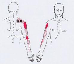
Deltoid
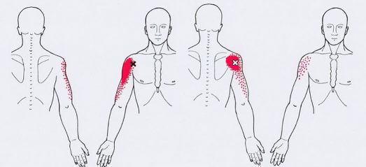
Pattern of referred pain and localization of trigger points in the right deltoid muscle. Left — a picture of pain from trigger points in the anterior muscles. On the right pictures — picture pain points in the posterior.
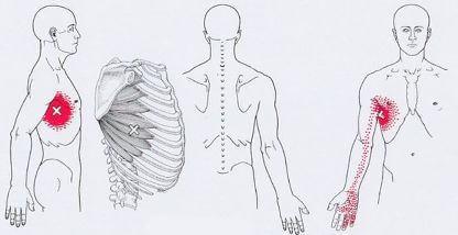
Coracobrachialis muscle
The picture of the pain and localization of trigger points in the right coracobrachialis muscle. Trigger points can be found in the distal or middle part of the muscle. Sometimes the pain from them applies only to the elbow.
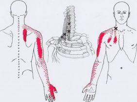
Two-headed muscle of the arm
Pattern of referred pain and localization of trigger points in the biceps muscle of the shoulder.
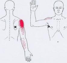
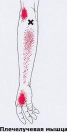
Shoulder muscle
Pattern of referred pain and localization of the trigger points in his right shoulder muscle. Please note: the upper trigger point can be a cause of compression of the radial nerve.
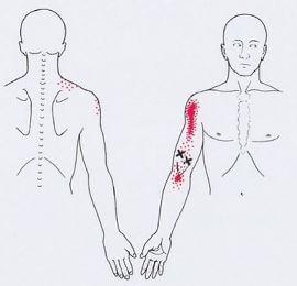
The triceps brachii
Pattern of referred pain and localization of trigger points in the triceps muscle of the shoulder. Left — ТТ1 in the left long head, ТТ2 in the lateral portion of the right middle head. In the centre — TTZ in the lateral edge of the lateral head, ТТ4 deeply in the distal right middle of head, center. Right -ТТ5 deep in the medial edge of the right medial heads.
Forearms and hands
Elbow muscle
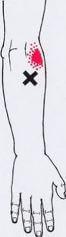
Localization of trigger points in the elbow muscle and the pattern of the reflected pain from them.
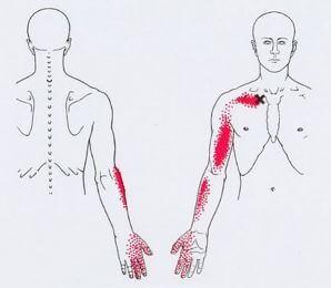
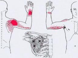
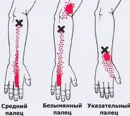
The extensor brush
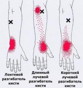
Pattern of referred pain and localization of trigger points in three main extensor of the brush on the right side.
Localization of trigger points in the right brachioradialis muscle and the pattern of the reflected pain from them.
The extensors of the fingers
Pattern of referred pain and localization of trigger points in the selected three muscles — the extensors of the fingers on his right hand.
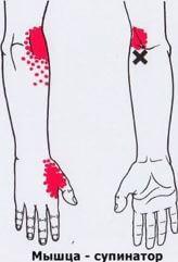
Localization of trigger points in the right supinator of the hand and the picture reflected her pain.
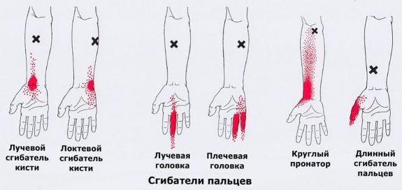
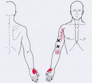
A composite picture of the reflected pain and localization of trigger points in the right flexors of the hand and fingers.
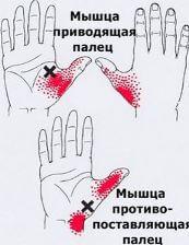
Pattern of referred pain and localization of trigger points in two muscles of the thumb of the right hand.
Pattern of referred pain and localization of trigger points in the intercostals muscles of the right hand. Trigger points can be found in any part of the interosseous spaces. Sometimes ihnasya nodules of Heberden.
Back and stomach
Surface okoloplodna muscles
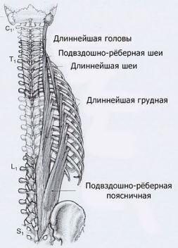
Attachment and the location of two of the most important surface groups of paravertebral muscles (the rectifier back).
Iliac rib thoracic
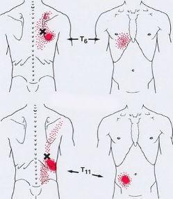
Pattern of referred pain and localization of trigger points in the thoracic iliac rib muscle.
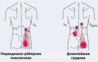
Pattern of referred pain and localization of trigger points in the lower thoracic and lumbar spine. Latin letters C, T, L, S, and the numbers indicate the levels of the vertebrae of the relevant departments.
Partitioned muscle
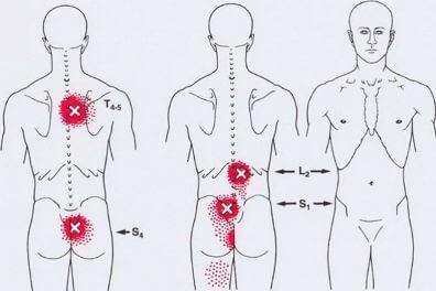
The picture reflected the pain and localization of trigger points in the deep paravertebral muscles ( partitioned and rotators). Left — example of trigger points in srednesrochnoi and niekraszewicz departments. Center and right — localization of TT in these muscles at the L2 and S1 vertebrae.
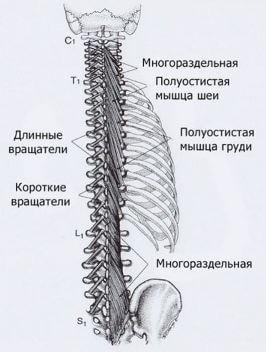
The attachment and location of the deep paravertebral muscles.
Partitioned the muscles of the neck
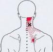
The picture reflected the pain and localization of trigger points in the deep muscles of the neck. Sometimes these points can cause compression of the great occipital nerve.
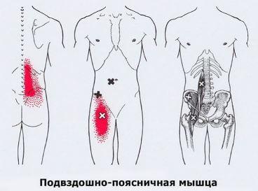
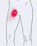
The picture reflected the pain and localization of trigger points in the right iliopsoas.
Obliques
Pattern of referred pain and visceral symptoms from trigger points, localized in the obliques (and possibly in the transverse mishey). Left — "heartburn" because of the trigger point of the external oblique muscle attaches to the anterior chest wall. Right groin pain and/or in the scrotum due to trigger points in the muscles of the lower lateral abdominal wall.
The rectus abdominis
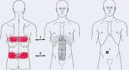
Pattern of referred pain and visceral symptoms due to trigger points in the straight muscle of the abdomen. Left and centre — bilateral pain across the back, feeling of overflow in the abdomen, nausea, vomiting can be caused by trigger points in the upper part of the rectus muscle. Similar bilateral pain in the lower divisions can be caused by points in zone 2.
The pelvis, buttocks and thighs.
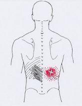
Square muscle of a loin
The picture reflected pain and trigger points in square loin muscle.
On the left and in the center of the marked trigger points, which can be palpated just below the 12 rib and above the Ilium. Right — trigger points in the deeper layers of the muscles.
The anal sphincter, the muscle that lifts the anus, PC muscle
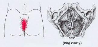
The internal obturator muscle
The picture reflected pain and trigger points in the pelvic floor muscles.
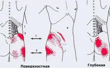
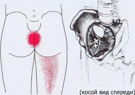
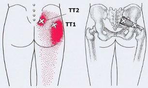
The gluteus Maximus
The picture reflected pain and trigger points in the gluteus Maximus muscle. Trigger points are localized: left (TT1), the upper-medial portion of the muscle. In the center (TT2) point in the region of the buttock. To the right is the medial lower portion (TTZ).
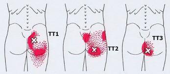
Gluteus Medius
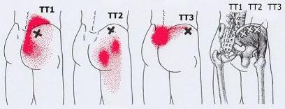
Pattern of referred pain from trigger points in the gluteal muscle harmful.
Medial point (ТТ1) reflect the pain in the iliac crest, in the region of the sacroiliac joint and the sacrum. ТТ2 located just above and lateral to and below reflect the pain in the buttocks. TTZ reflects bilateral pain in the rump and nizhnespasskoe Department.
Small gluteal muscle
In the figures, the pattern of referred pain from trigger points in the anterior portion of a small right gluteal muscle.
More areas appear full on muscle work. On the right drawings, the point in the front portion of the muscle.
The piriformis muscle
The overall picture of pain from trigger points in the right piriformis muscle. The most frequent lateral point (ТТ1)
Hip and knees
The tensioner broad fascia of the thigh
Pattern of referred pain from trigger points in the right muscle tensioning broad fascia of the thigh. Fascia figure removed.
The Sartorius muscle
Referred pain from the three trigger points in the right Sartorius muscle at different levels. Front-side view. Trigger points in this long muscle located superficially, sometimes right under the skin.
Scalloping the muscle
In the Pattern of referred pain from trigger points in the right pectinate muscle.
Quadriceps femoris
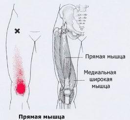
Pattern of referred pain from trigger points in the right direct muscle of the thigh.
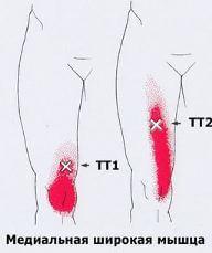
Pattern of referred pain from trigger points in the right medial broad the thigh muscle.
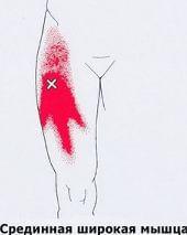
Pattern of referred pain from trigger points in the right middle of the broad muscle of the thigh.
In the distal may be an additional trigger points.
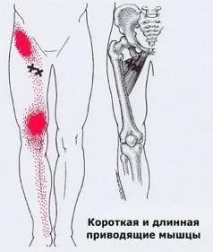
Front view on the right muscles, leading thigh. The picture of the reflected pain from trigger points in these muscles.
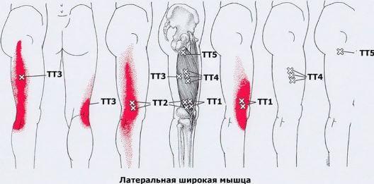
Pattern of referred pain from trigger points in the right lateral wide thigh muscle. Also shown is the straight muscle of the thigh.
Point TT1 can block the mobility of the patella. ТТ4 "closes" the broad fascia of the thigh and causes a sharp pain, does not sleep on the affected side.
Slender muscle
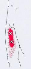
Front-side view of the overall picture of pain reflected from trigger points in the right slender muscle.
A large adductor muscle
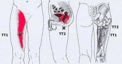
Pattern of referred pain from trigger points in the right is a big muscle that causes hip.
Left — front view. In the centre is shown the pain in the inside of the pelvis, caused by points within the area ТТ2. These points lie deep and sometimes located beneath the large gluteal muscles. To the right anatomical illustration.
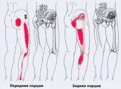
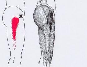
The muscles back of the thigh
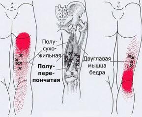
Pattern of referred pain from trigger points in the muscles of the back of the thigh.
Popliteal muscle
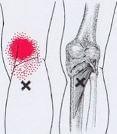
Pattern of referred pain from trigger points in the right popliteal, right mice
It must be remembered that in this area are the major arteries, veins and nerves that can be affected.
Lower leg, ankle and foot
Anterior tibial muscle
Pattern of referred pain and localization of trigger points in the right tibialis anterior muscle.
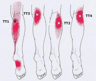
Long extensor digitorum
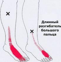
Pattern of referred pain and localization of trigger points in the right muscles extending the toes.
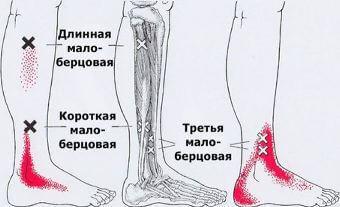
Pattern of referred pain from trigger points in the peroneal muscles of the right leg.
It is seen that almost all points reflect the pain in the distal zone of the leg and foot.
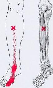
Calf
Localization of trigger points in the right gastrocnemius muscle and the pattern of reflected pain. ТТ1 is the medial head, ТТ2 — lateral. These points can cause night cramps — "crampy". Two more proksimalna points TTZ иТТ4 projecting pain under the knee.
Pattern of referred pain from trigger points, localized on the short flexors of the fingers. Solid red shows the basic area of pain, granular -additional zone.
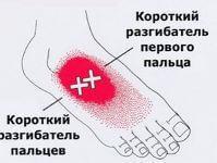
Muscle, abductor of the first finger
Pattern of referred pain from trigger points, localized in the muscle discharge of the first finger.
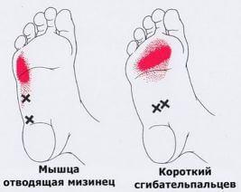
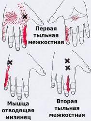
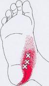
Pattern of referred pain from trigger points, localized in the two superficial muscles of the right foot: short flexor of the fingers and the abductor little finger.
Square muscle of the foot
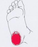
Pattern of referred pain from trigger points, localized to deep square muscle of the foot. This pain may mimic the presence of a "heel spur".
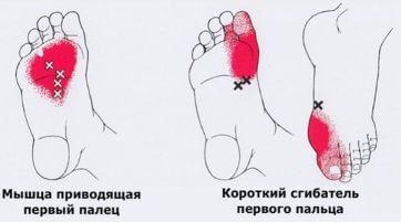
Pattern of referred pain from trigger points, localized in two deep internal muscles of the first toe of the right foot: flexor and adductor muscles.
The first interosseous muscle
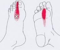
A typical picture of pain reflected from trigger points in the first interosseous muscle of the right foot. published
P. S. And remember, only by changing their consumption — together we change the world! ©
Source: svoistva-tela.ru/html/tochki-boli-i-napryazheniya.html
Solid red shows the basic area of pain, granular - additional zone.
Crosses marked trigger points (stress points).
Head and neck
Trapezoidal muscle

Pattern of referred pain and localization of trigger points in the upper part of trapeze muscle.
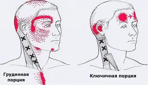
Sternocleidomastoid muscle

Pattern of referred pain and localization of the responsible trigger points in the right sternocleidomastoid muscle. On the left of the sternum (surface) portion. Right — clavicular (deep) portion.
The masseter

Localization of trigger points in various parts of the chewing muscles. Solid red shows the basic area of pain, granular — additional zone. Left — surface layer, the upper and middle division. In the center — superficial layer, lower part. Right — deep layer, upper part, just below the temporomandibular joint.
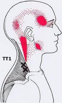
The temporalis muscle

The picture of the reflected pain from TRIG-guérin dots in the left temporal muscle. Solid red shows the basic area of pain, granular — additional zone. Front "needle" pain arises from the anterior fibers (ТТ1), the middle "spokes" of ТТ2 and TTZ, rear (nadolna) "spoke" of ТТ4.
Medial hyoid muscle

Picture otrajenii pain (marked in red) and localization of the responsible trigger points in the medial hyoid muscle. Left — the outer area of pain, which can indicate patients. Right — the picture of the internal part of the pain was passing through the temporomandibular joint.
The lateral hyoid muscle

Pattern of referred pain from trigger points in the lateral hyoid muscle.
Digastric

Picture of trigger points, and reflected from them, pain in the right dobrydney muscle.
Left — back abdomen — side view. Right — front abdomen — front view.
Suboccipital muscle

The picture reflected pain and trigger points in the right suboccipital muscle.

Pattern of referred pain (marked in red) and location of trigger points in the muscles polyostotic. Left upper trigger point in the muscle polyostotic head. To the right is the trigger point in the third layer polyostotic muscles of the neck.

Trigger points and pattern they cause pain in the right belt muscle of the head and neck. On the left drawings — trigger points in the belt muscles of the head, in the occipital triangle. The figures on the right — upper trigger point that causes pain in the eye orbits, the lower the trigger point that causes pain in the angle of the neck.
Shoulders, chest and arms
Trapezoidal muscle

Referred pain and localization ТТ2 in the upper part of trapeze muscle, TTZ, ТТ4 at the bottom, ТТ5, TTB — in secondary departments trapezoidal muscles.
Muscle, levator scapulae

Picture of mixed pain, caused by two trigger points in the right muscle, levator scapulae. Solid red shows the basic area of pain, granular — additional zone.
Scalene muscle

A complex picture of pain caused by trigger points located in the anterior, middle and posterior scalene muscles. Some trigger points may have only one ongoing area otrajenii pain.
The pectoralis major muscle

Pattern of referred pain and localization of trigger points in the pectoralis major muscle.
On the leftmost figure shows how overlapped the pain referred from the two trigger points located in the middle part of the muscle around of the sternum. Further, the figures show: left — the trigger point in the intermediate part Grudin-tion division, in the center — of TT in the clavicular part to the right is the trigger point of the free edges of the muscle that forms the axilla.

Trigger points in the right pectoralis minor muscle and its pain pattern caused.

Pattern of referred pain caused by trigger point in the left breast muscle.
Subclavian muscle

Pattern of referred pain caused by trigger point in the right subclavian muscle.
The serratus anterior muscle

Pattern of referred pain caused by trigger point localized in the right serratus anterior. Side view, back and front.
Rear-upper serrated muscle

Pattern of referred pain from trigger points in the right-rear-upper serrated muscle. Areas of persistent pain is marked by a solid red. Granular marked areas of possible pain. At left — back. The figure in the center of the blade assigned forward and the trigger point is available for palpation and injection. On the right picture the front view.
Rear-lower serrated muscle

Pattern of referred pain from trigger points in the right-rear-lower serrated muscle.
Latissimus dorsi

Pattern of referred pain and trigger points in the right-the widest muscle in the back. To the left is the usual localization of trigger points in the axillary portions of the muscle. In the center — front view. Right — the picture of pain from the lower trigger point.
Supraspinatus muscle

Pattern of referred pain and localization of trigger points in the right supraspinatus muscle.
Infraspinatus muscle

The picture reflected the pain and localization of trigger points in the right infraspinatus muscle.
Small round muscle

Pattern of referred pain and localization of trigger points in the right of the small round muscle.
Large round muscle

Medial and lateral (back and axillary) trigger points in the right a large circular muscle and the pattern of reflected pain. Left — medial trigger points, the right — lateral TT.
Subscapularis muscle

Pattern of referred pain from trigger points in the right subscapularis muscle.
Rhomboid muscle

The overall picture of pain from trigger points in the right rhomboid muscle.
Deltoid

Pattern of referred pain and localization of trigger points in the right deltoid muscle. Left — a picture of pain from trigger points in the anterior muscles. On the right pictures — picture pain points in the posterior.
Coracobrachialis muscle

The picture of the pain and localization of trigger points in the right coracobrachialis muscle. Trigger points can be found in the distal or middle part of the muscle. Sometimes the pain from them applies only to the elbow.
Two-headed muscle of the arm

Pattern of referred pain and localization of trigger points in the biceps muscle of the shoulder.
Shoulder muscle

Pattern of referred pain and localization of the trigger points in his right shoulder muscle. Please note: the upper trigger point can be a cause of compression of the radial nerve.
The triceps brachii

Pattern of referred pain and localization of trigger points in the triceps muscle of the shoulder. Left — ТТ1 in the left long head, ТТ2 in the lateral portion of the right middle head. In the centre — TTZ in the lateral edge of the lateral head, ТТ4 deeply in the distal right middle of head, center. Right -ТТ5 deep in the medial edge of the right medial heads.
Forearms and hands
Elbow muscle

Localization of trigger points in the elbow muscle and the pattern of the reflected pain from them.
The extensor brush

Pattern of referred pain and localization of trigger points in three main extensor of the brush on the right side.

Localization of trigger points in the right brachioradialis muscle and the pattern of the reflected pain from them.
The extensors of the fingers

Pattern of referred pain and localization of trigger points in the selected three muscles — the extensors of the fingers on his right hand.

Localization of trigger points in the right supinator of the hand and the picture reflected her pain.

A composite picture of the reflected pain and localization of trigger points in the right flexors of the hand and fingers.

Pattern of referred pain and localization of trigger points in two muscles of the thumb of the right hand.

Pattern of referred pain and localization of trigger points in the intercostals muscles of the right hand. Trigger points can be found in any part of the interosseous spaces. Sometimes ihnasya nodules of Heberden.
Back and stomach
Surface okoloplodna muscles

Attachment and the location of two of the most important surface groups of paravertebral muscles (the rectifier back).
Iliac rib thoracic

Pattern of referred pain and localization of trigger points in the thoracic iliac rib muscle.

Pattern of referred pain and localization of trigger points in the lower thoracic and lumbar spine. Latin letters C, T, L, S, and the numbers indicate the levels of the vertebrae of the relevant departments.
Partitioned muscle

The picture reflected the pain and localization of trigger points in the deep paravertebral muscles ( partitioned and rotators). Left — example of trigger points in srednesrochnoi and niekraszewicz departments. Center and right — localization of TT in these muscles at the L2 and S1 vertebrae.

The attachment and location of the deep paravertebral muscles.
Partitioned the muscles of the neck

The picture reflected the pain and localization of trigger points in the deep muscles of the neck. Sometimes these points can cause compression of the great occipital nerve.

The picture reflected the pain and localization of trigger points in the right iliopsoas.
Obliques

Pattern of referred pain and visceral symptoms from trigger points, localized in the obliques (and possibly in the transverse mishey). Left — "heartburn" because of the trigger point of the external oblique muscle attaches to the anterior chest wall. Right groin pain and/or in the scrotum due to trigger points in the muscles of the lower lateral abdominal wall.
The rectus abdominis

Pattern of referred pain and visceral symptoms due to trigger points in the straight muscle of the abdomen. Left and centre — bilateral pain across the back, feeling of overflow in the abdomen, nausea, vomiting can be caused by trigger points in the upper part of the rectus muscle. Similar bilateral pain in the lower divisions can be caused by points in zone 2.
The pelvis, buttocks and thighs.
Square muscle of a loin

The picture reflected pain and trigger points in square loin muscle.
On the left and in the center of the marked trigger points, which can be palpated just below the 12 rib and above the Ilium. Right — trigger points in the deeper layers of the muscles.
The anal sphincter, the muscle that lifts the anus, PC muscle

The internal obturator muscle

The picture reflected pain and trigger points in the pelvic floor muscles.
The gluteus Maximus

The picture reflected pain and trigger points in the gluteus Maximus muscle. Trigger points are localized: left (TT1), the upper-medial portion of the muscle. In the center (TT2) point in the region of the buttock. To the right is the medial lower portion (TTZ).
Gluteus Medius

Pattern of referred pain from trigger points in the gluteal muscle harmful.
Medial point (ТТ1) reflect the pain in the iliac crest, in the region of the sacroiliac joint and the sacrum. ТТ2 located just above and lateral to and below reflect the pain in the buttocks. TTZ reflects bilateral pain in the rump and nizhnespasskoe Department.
Small gluteal muscle

In the figures, the pattern of referred pain from trigger points in the anterior portion of a small right gluteal muscle.
More areas appear full on muscle work. On the right drawings, the point in the front portion of the muscle.
The piriformis muscle

The overall picture of pain from trigger points in the right piriformis muscle. The most frequent lateral point (ТТ1)
Hip and knees
The tensioner broad fascia of the thigh

Pattern of referred pain from trigger points in the right muscle tensioning broad fascia of the thigh. Fascia figure removed.
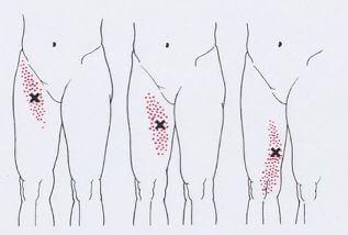
The Sartorius muscle

Referred pain from the three trigger points in the right Sartorius muscle at different levels. Front-side view. Trigger points in this long muscle located superficially, sometimes right under the skin.
Scalloping the muscle

In the Pattern of referred pain from trigger points in the right pectinate muscle.
Quadriceps femoris

Pattern of referred pain from trigger points in the right direct muscle of the thigh.

Pattern of referred pain from trigger points in the right medial broad the thigh muscle.

Pattern of referred pain from trigger points in the right middle of the broad muscle of the thigh.
In the distal may be an additional trigger points.

Front view on the right muscles, leading thigh. The picture of the reflected pain from trigger points in these muscles.

Pattern of referred pain from trigger points in the right lateral wide thigh muscle. Also shown is the straight muscle of the thigh.
Point TT1 can block the mobility of the patella. ТТ4 "closes" the broad fascia of the thigh and causes a sharp pain, does not sleep on the affected side.
Slender muscle

Front-side view of the overall picture of pain reflected from trigger points in the right slender muscle.
A large adductor muscle

Pattern of referred pain from trigger points in the right is a big muscle that causes hip.
Left — front view. In the centre is shown the pain in the inside of the pelvis, caused by points within the area ТТ2. These points lie deep and sometimes located beneath the large gluteal muscles. To the right anatomical illustration.
The muscles back of the thigh

Pattern of referred pain from trigger points in the muscles of the back of the thigh.
Popliteal muscle

Pattern of referred pain from trigger points in the right popliteal, right mice
It must be remembered that in this area are the major arteries, veins and nerves that can be affected.
Lower leg, ankle and foot
Anterior tibial muscle

Pattern of referred pain and localization of trigger points in the right tibialis anterior muscle.
Long extensor digitorum

Pattern of referred pain and localization of trigger points in the right muscles extending the toes.

Pattern of referred pain from trigger points in the peroneal muscles of the right leg.
It is seen that almost all points reflect the pain in the distal zone of the leg and foot.
Calf

Localization of trigger points in the right gastrocnemius muscle and the pattern of reflected pain. ТТ1 is the medial head, ТТ2 — lateral. These points can cause night cramps — "crampy". Two more proksimalna points TTZ иТТ4 projecting pain under the knee.

Pattern of referred pain from trigger points, localized on the short flexors of the fingers. Solid red shows the basic area of pain, granular -additional zone.
Muscle, abductor of the first finger

Pattern of referred pain from trigger points, localized in the muscle discharge of the first finger.

Pattern of referred pain from trigger points, localized in the two superficial muscles of the right foot: short flexor of the fingers and the abductor little finger.
Square muscle of the foot

Pattern of referred pain from trigger points, localized to deep square muscle of the foot. This pain may mimic the presence of a "heel spur".

Pattern of referred pain from trigger points, localized in two deep internal muscles of the first toe of the right foot: flexor and adductor muscles.
The first interosseous muscle

A typical picture of pain reflected from trigger points in the first interosseous muscle of the right foot. published
P. S. And remember, only by changing their consumption — together we change the world! ©
Source: svoistva-tela.ru/html/tochki-boli-i-napryazheniya.html
Tags
See also
A simple technique Haruki Nakamura to relieve tension in the neck muscles and improve blood circulation
What we know about ourselves ...
5 secrets of the human race, that science still can not explain
The post on the water and 4 of the method of detoxification of the body
Modern technologies for the benefit of
Excursion to the biggest fridge in Ukraine.
Pony apocalypse
121 tips on how to uncover possible brain
Sea lions have a sense of rhythm
My name is Red