Unique coverage of the female body - the story of the origin of life
 Bashny.Net
We offer you the unique coverage of the female body.
These unique photographs taken by the Swedish photographer Lennart Nilsson back in 1965. The photographer was born in 1922 and became the first person with the help of special equipment and cameras, was able to get inside a woman's body and remove the whole process of the birth of a new life.
First pictures of the embryo appeared in print as early as 1953, and this event inspired the photographer to create new jobs.
In order to show the development of man from the beginning, and he placed the microchamber mikroosvetitel at the end of the tube cystoscope, which examine the bladder and removed his unique photos directly from where people take the first step into the world of ...
Here it is - our history is the origin of life!
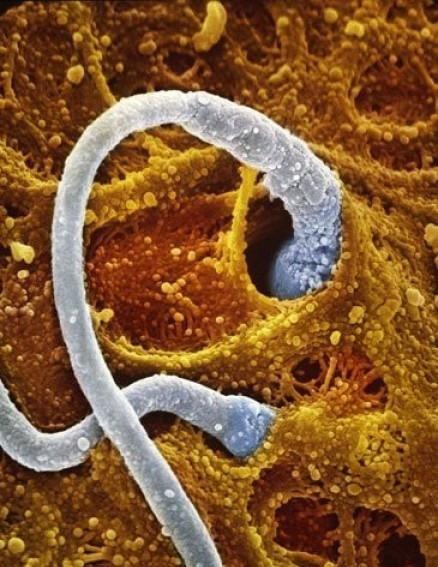
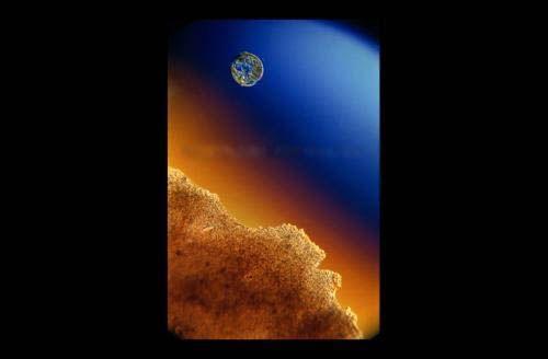
Here the sperm moves toward the egg
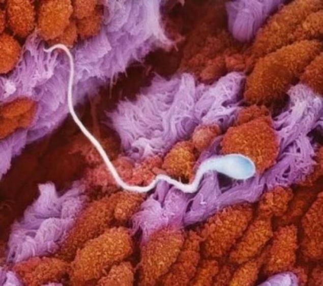
Sperm in the folds of the mucous membrane of the fallopian tubes is moving toward the egg.
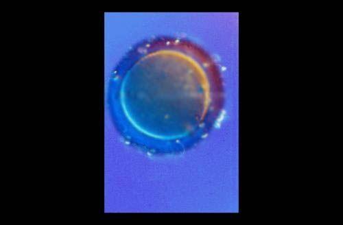
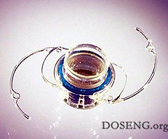
The egg ...
Will there be a meeting?
The walls of the fallopian tubes ...
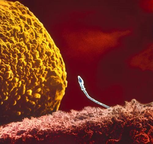
Two sperm in contact with the shell of the egg. Enzymes contained in the head of the sperm, the egg shell was dissolved, but the genetic material of the sperm is involved in fertilization.
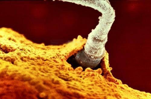
One of the father's sperm 200 million, breaking through the shell of the egg, just poured into it ...
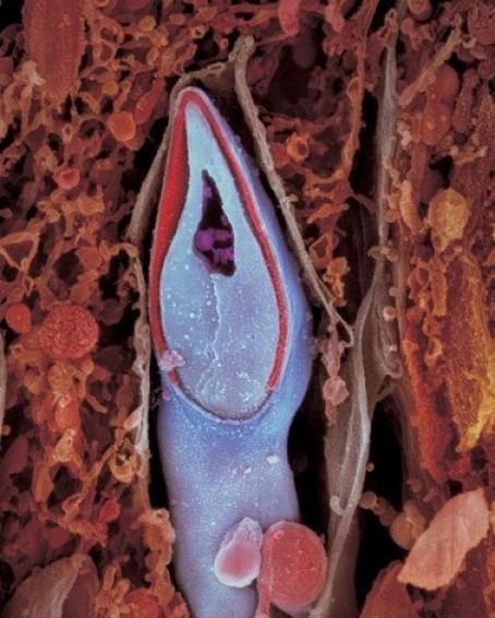
Longitudinal section of the sperm. The genetic material contained in the sperm head
A week later, the embryo, sliding down the fallopian tube into the uterus moves ...
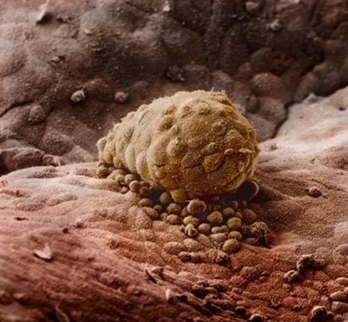
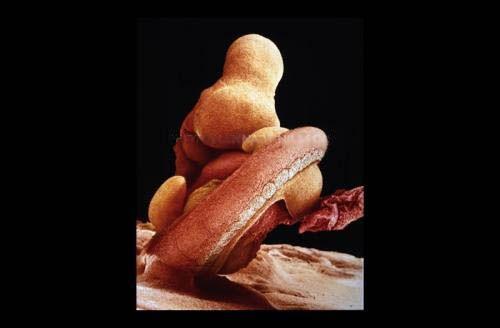
The embryo, attached to the endometrium (8 days)
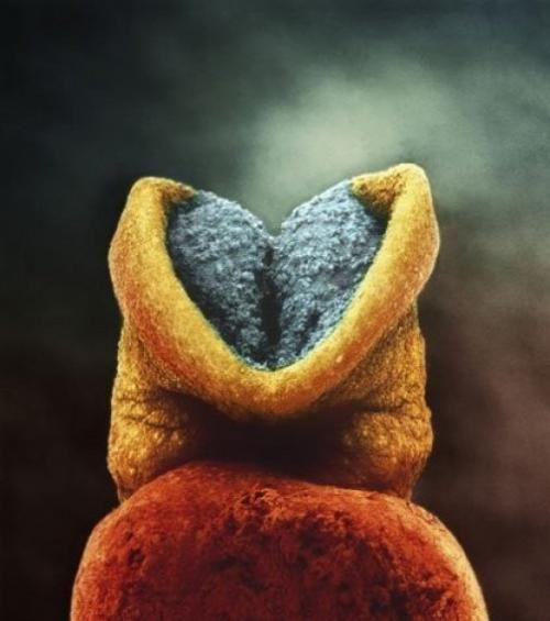
The development of the embryo. Grey color - future brain. (22 days)
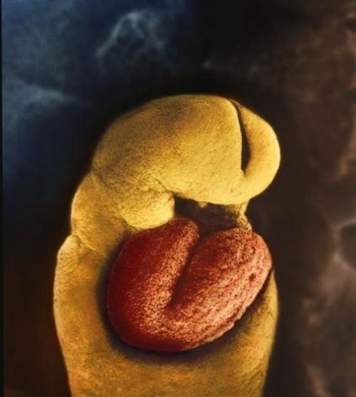
24 days. Skeleton at the month of the embryo are no - there is only the heart, it begins to pulsate on the 18th day
The 28th day after fertilization ...
4 and a half weeks ...
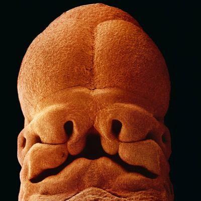
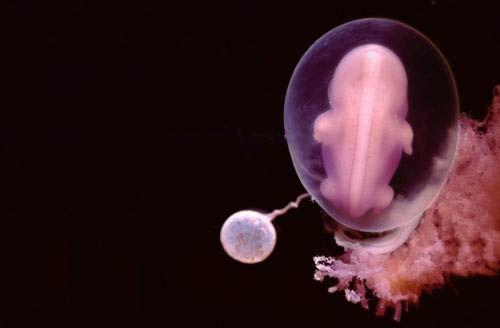
Five-week embryo, length 9 mm, have guessed the face with holes for mouth, nostrils and eyes.
40 days. Outside the cell nucleus fused with a loose surface of the uterus and form the placenta or afterbirth. It is a spongy piece of flesh for a man in the first nine months of his life and light, and the stomach and the liver, and kidney ...
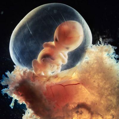
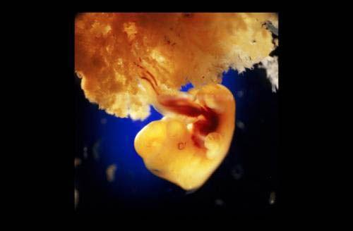
Eight weeks. Rapidly growing embryo is well protected in the womb. With the help of an electron microscope Nilsson was able to increase the image of hundreds of thousands of times.
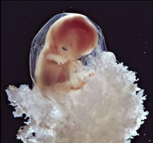
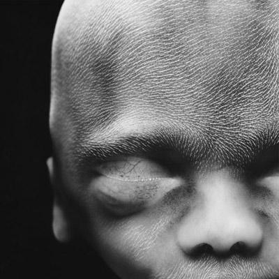
10 weeks. Eyelids are already half open. Within a few days, they will form completely.
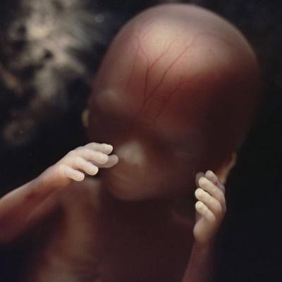
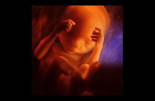
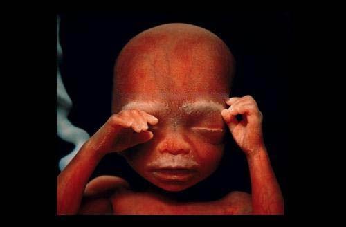
16 weeks. An inquisitive kid is already using his hands to explore the area.
The skeleton is mainly composed of a flexible rod and a network of blood vessels visible through the thin skin.
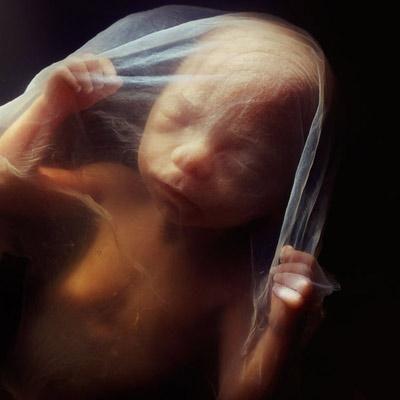
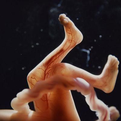
18 weeks. About 14 cm. The fetus can now perceive sounds from the outside world.

19 weeks ...
20 weeks. About 20 cm. On the head the hair is already starting to appear.
24 weeks ...
26 weeks ...
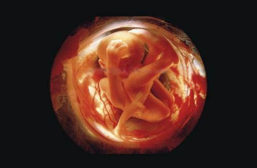
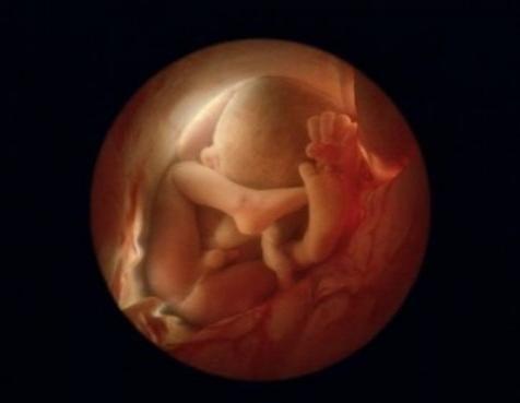
6 months. There are still eight to ten weeks of carefree, but the little man already crowded in the uterus, and he is preparing to leave her. He turns upside down - it's more convenient to get ...
36 weeks. After 4 weeks, the child sees the white light
Source: imarocksta.livejournal.com
These unique photographs taken by the Swedish photographer Lennart Nilsson back in 1965. The photographer was born in 1922 and became the first person with the help of special equipment and cameras, was able to get inside a woman's body and remove the whole process of the birth of a new life.
First pictures of the embryo appeared in print as early as 1953, and this event inspired the photographer to create new jobs.
In order to show the development of man from the beginning, and he placed the microchamber mikroosvetitel at the end of the tube cystoscope, which examine the bladder and removed his unique photos directly from where people take the first step into the world of ...
Here it is - our history is the origin of life!
Here the sperm moves toward the egg

Sperm in the folds of the mucous membrane of the fallopian tubes is moving toward the egg.

The egg ...

Will there be a meeting?

The walls of the fallopian tubes ...

Two sperm in contact with the shell of the egg. Enzymes contained in the head of the sperm, the egg shell was dissolved, but the genetic material of the sperm is involved in fertilization.

One of the father's sperm 200 million, breaking through the shell of the egg, just poured into it ...

Longitudinal section of the sperm. The genetic material contained in the sperm head

A week later, the embryo, sliding down the fallopian tube into the uterus moves ...

The embryo, attached to the endometrium (8 days)

The development of the embryo. Grey color - future brain. (22 days)

24 days. Skeleton at the month of the embryo are no - there is only the heart, it begins to pulsate on the 18th day

The 28th day after fertilization ...

4 and a half weeks ...

Five-week embryo, length 9 mm, have guessed the face with holes for mouth, nostrils and eyes.

40 days. Outside the cell nucleus fused with a loose surface of the uterus and form the placenta or afterbirth. It is a spongy piece of flesh for a man in the first nine months of his life and light, and the stomach and the liver, and kidney ...

Eight weeks. Rapidly growing embryo is well protected in the womb. With the help of an electron microscope Nilsson was able to increase the image of hundreds of thousands of times.

10 weeks. Eyelids are already half open. Within a few days, they will form completely.

16 weeks. An inquisitive kid is already using his hands to explore the area.

The skeleton is mainly composed of a flexible rod and a network of blood vessels visible through the thin skin.

18 weeks. About 14 cm. The fetus can now perceive sounds from the outside world.

19 weeks ...

20 weeks. About 20 cm. On the head the hair is already starting to appear.

24 weeks ...

26 weeks ...

6 months. There are still eight to ten weeks of carefree, but the little man already crowded in the uterus, and he is preparing to leave her. He turns upside down - it's more convenient to get ...

36 weeks. After 4 weeks, the child sees the white light
Source: imarocksta.livejournal.com
Tags
See also
25 unique photos of National Geographic, previously unpublished
The history of the life of giant pandas
Nepridumannoe GOOD STORIES FROM THE LIVES
Unique photos 2
The history of life Misha
The sad story of the life of a cat ... with a happy ending!
Bacteria fix atmospheric nitrogen learned a billion years earlier than previously thought
IBM-PC-incompatible shooters
Overheard on the Internet: the real stories from life, from which you will laugh that is forces.
Speaking of luxury brains ... or ... 10 ways to sell anything ...