The best pictures from the world of medicine and biology in 2009
 Bashny.Net
In continuation of the mega-popular theme
Micro-images from the depths of the human body
Is the world of medicine can be truly beautiful? It turns out, can, particularly when viewed through an electron microscope. UK's largest charitable foundation Wellcome Trust this year already holds a competition of the best photos related to the field of medicine, biology and health care. Today we suggest to familiarize with the best pictures, the winners in 2009.
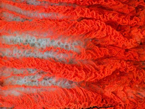
Photo sterlittsii grains or paradise flower Strelitzia reginae (© Annie Cavanagh & Dave McCarthy). Made with an electron microscope. Homeland of the plant is South Africa. Features sterlittsii - orange-blue color of the flower.
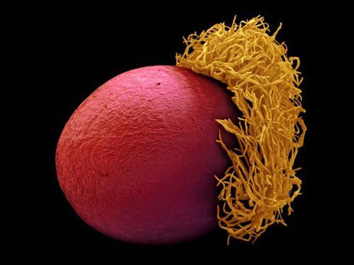
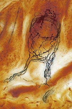
Photo finest blood vessels of the ciliary body of the bull's eye (© MI Walker). Done using an optical microscope. According to the author, picture configured of 27 shots that allowed us to obtain a three-dimensional effect. Capillaries became visible only after injection of the insoluble dye into an artery.
In the picture below shows the nerve endings at the end of the hair follicle (© MI Walker). Black color have axons.
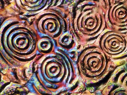
It turns out that man-made substances and objects under a microscope can also look amazing. Before you - aspirin crystals photograph taken with an optical microscope (© MI Walker).
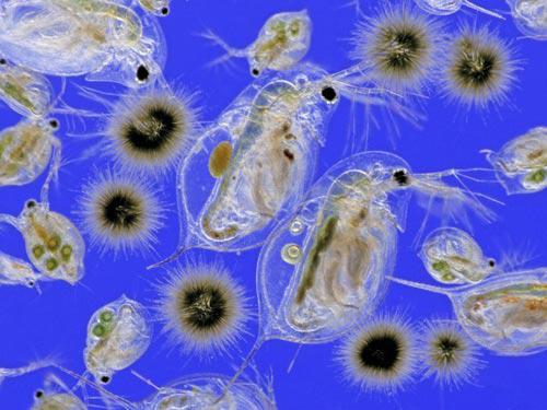
And it is - an ordinary plankton (© MI Walker). For this image the author had to resort to the use of lighting techniques Rheinberg (Rheinberg illumination). Recall that the plankton inhabit the waters of the seas and oceans and is divided into two large groups or categories - phytoplankton and zooplankton.
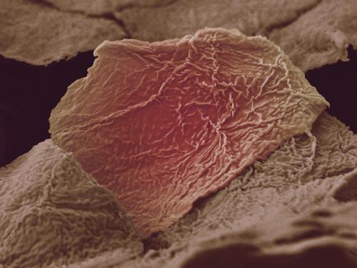
The object of study in this picture - burns caused by contact with skin hot soup (© Anne Weston). The damaged area was photographed using a scanning electron microscope (SEM). Interestingly, the author of the picture was the victim herself - Annie Weston.
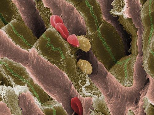
In this photo you can see the internal structure of mouse liver (© Jackie Lewin).
The next step - a three-dimensional model of the head of a mouse embryo at the stage (© NIMR, MRC). Obtained by epi high-resolution microscopy (high-resolution episcopic microscopy, HREM). The computer scientists were able to "connect" head shots and click to create a three-dimensional figure.
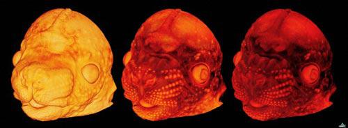
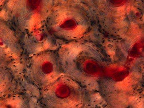
Photo of the human hip bone compact (© Ivor Mason). In the picture you can see the finest blood vessels, connective tissue. The structure of compact bone material includes organic and inorganic salts.
via
Source:
Micro-images from the depths of the human body
Is the world of medicine can be truly beautiful? It turns out, can, particularly when viewed through an electron microscope. UK's largest charitable foundation Wellcome Trust this year already holds a competition of the best photos related to the field of medicine, biology and health care. Today we suggest to familiarize with the best pictures, the winners in 2009.
Photo sterlittsii grains or paradise flower Strelitzia reginae (© Annie Cavanagh & Dave McCarthy). Made with an electron microscope. Homeland of the plant is South Africa. Features sterlittsii - orange-blue color of the flower.

Photo finest blood vessels of the ciliary body of the bull's eye (© MI Walker). Done using an optical microscope. According to the author, picture configured of 27 shots that allowed us to obtain a three-dimensional effect. Capillaries became visible only after injection of the insoluble dye into an artery.

In the picture below shows the nerve endings at the end of the hair follicle (© MI Walker). Black color have axons.

It turns out that man-made substances and objects under a microscope can also look amazing. Before you - aspirin crystals photograph taken with an optical microscope (© MI Walker).

And it is - an ordinary plankton (© MI Walker). For this image the author had to resort to the use of lighting techniques Rheinberg (Rheinberg illumination). Recall that the plankton inhabit the waters of the seas and oceans and is divided into two large groups or categories - phytoplankton and zooplankton.

The object of study in this picture - burns caused by contact with skin hot soup (© Anne Weston). The damaged area was photographed using a scanning electron microscope (SEM). Interestingly, the author of the picture was the victim herself - Annie Weston.

In this photo you can see the internal structure of mouse liver (© Jackie Lewin).

The next step - a three-dimensional model of the head of a mouse embryo at the stage (© NIMR, MRC). Obtained by epi high-resolution microscopy (high-resolution episcopic microscopy, HREM). The computer scientists were able to "connect" head shots and click to create a three-dimensional figure.

Photo of the human hip bone compact (© Ivor Mason). In the picture you can see the finest blood vessels, connective tissue. The structure of compact bone material includes organic and inorganic salts.
via

Source:
Tags
See also
Comedy Wildlife Photography Awards 2015: best pictures of the funny photo contest in the world
The best pictures of weather phenomena from NASA
Experts have called the best job in the world
Best father in the world
10 of the best spa resorts in the world
The best pictures in 2011. Part 3
The best pictures in 2011. Part 2
The best father in the world
15 best pictures of tornado season in 2013
Top 25 pictures of Jan Arthus Bertrand (25 photos)