Fig- contest
 Bashny.Net
Remember the very popular theme "Micro-images from the depths of the human body»?
At the competition «Olympus BioScapes Imaging 2009" presents microscopic photographs of objects of biological science. Presenting your attention the photos of ten winners of the competition this year.
1. In this photo taken biologist David Skidmore College Domozychem you see unicellular alga gene that breaks down after contact with poisonous microtubules known as oryzalin. This photo took fifth place in the competition «Olympus BioScapes Digital Imaging 2009". (David Domozych)

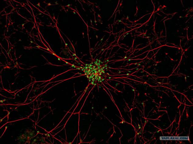
2. This picture Gist Croft and Mackenzie Veygrandt Columbia University took 10th place in the competition. The images show the motor neuron, struck amyotrophic lateral sclerosis, also known as Lou Gehrig's disease. For this picture skin cells were taken 83-year-old patient with amyotrophic lateral sclerosis and reprogrammed into induced pluripotent stem cells. Then the cells have been transformed into neurons. The study of these neurons will help scientists to confront amyotrophic lateral sclerosis. (Gist Croft and Mackenzie Weygandt)
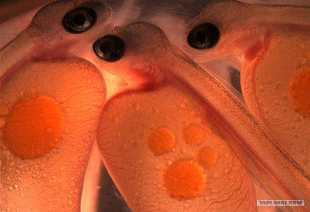
3. Haruki Fudzhimake Mount Holyoke college in Massachusetsve made this vivid picture of embryos of Atlantic salmon from Bryant Pond, Maine. Photo took ninth place in the competition «Olympus BioScapes Digital Imaging 2009". (Haruka Fujimaki)
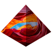
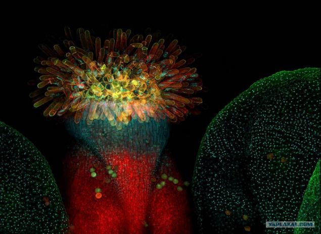
4. Chait Peyvs - researcher at the Tallinn University of Technology - used confocal microscopy to create this unusual image of the flower of Arabidopsis, which is a common model organism in plant biology and genetic engineering. This photo took eighth place in the competition «Olympus BioScapes Digital Imaging 2009". (Heiti Paves)
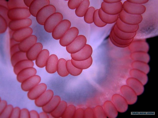
5. notoriously painful powerful uzhalivaniem, Portuguese ship is a floating cavity filled with gas that supports the tentacles with stinging cells. In this picture Alvaro Migotto from the University of Sao Paulo you see pink "charges" stinging cells, and a thin strand of muscle, which is responsible for high contractility tentacles. The picture won second place. (Alvaro Migotto)
7. This picture, the runner-up, was made Chun-Ju Rachel Wong of Berkeley. On it you can see the step the protein structure inside the nucleus of the plant cell corn. The structure, known as synaptonemal complex formed between the paired chromosomes during cell division. This may be the first three-dimensional image with high resolution ever done by optical microscopy. Two parallel axes set extending along the entire length of the chromosomes, the two strands are at a distance of 100 nm from one another and enveloping each other in a spiral. (Chung-Ju Rachel Wang)

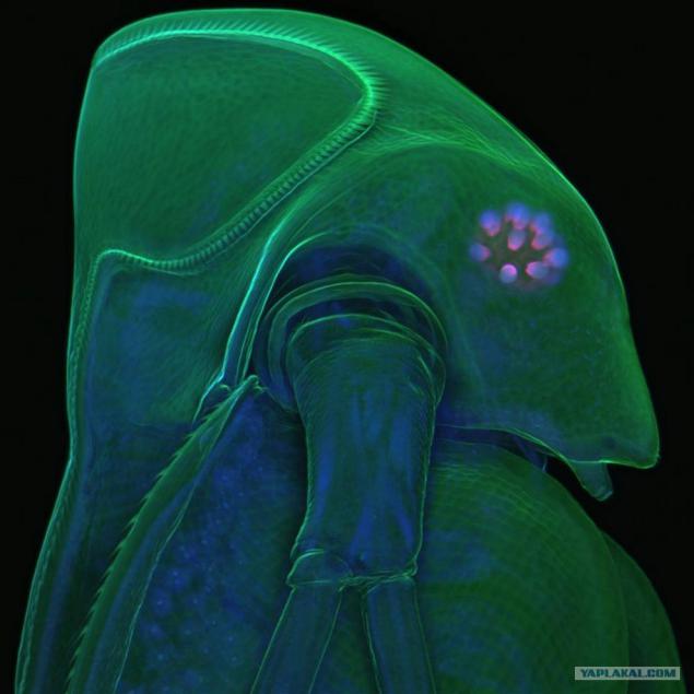
8. This spooky and at the same time a beautiful picture of the water with his bi bright green "crown of thorns" won the contest «Olympus BioScapes Digital Imaging 2009". "Crown" exoskeleton protects goods from predators. This picture taken by a zoologist at the University of Jan Michels Albrecht, reveals not only the exoskeleton, but also the internal parts - right down to the core cells being, which can be seen here in the form of tiny twinkling dots. (Jan Michels)
Source:
At the competition «Olympus BioScapes Imaging 2009" presents microscopic photographs of objects of biological science. Presenting your attention the photos of ten winners of the competition this year.
1. In this photo taken biologist David Skidmore College Domozychem you see unicellular alga gene that breaks down after contact with poisonous microtubules known as oryzalin. This photo took fifth place in the competition «Olympus BioScapes Digital Imaging 2009". (David Domozych)

2. This picture Gist Croft and Mackenzie Veygrandt Columbia University took 10th place in the competition. The images show the motor neuron, struck amyotrophic lateral sclerosis, also known as Lou Gehrig's disease. For this picture skin cells were taken 83-year-old patient with amyotrophic lateral sclerosis and reprogrammed into induced pluripotent stem cells. Then the cells have been transformed into neurons. The study of these neurons will help scientists to confront amyotrophic lateral sclerosis. (Gist Croft and Mackenzie Weygandt)

3. Haruki Fudzhimake Mount Holyoke college in Massachusetsve made this vivid picture of embryos of Atlantic salmon from Bryant Pond, Maine. Photo took ninth place in the competition «Olympus BioScapes Digital Imaging 2009". (Haruka Fujimaki)

4. Chait Peyvs - researcher at the Tallinn University of Technology - used confocal microscopy to create this unusual image of the flower of Arabidopsis, which is a common model organism in plant biology and genetic engineering. This photo took eighth place in the competition «Olympus BioScapes Digital Imaging 2009". (Heiti Paves)

5. notoriously painful powerful uzhalivaniem, Portuguese ship is a floating cavity filled with gas that supports the tentacles with stinging cells. In this picture Alvaro Migotto from the University of Sao Paulo you see pink "charges" stinging cells, and a thin strand of muscle, which is responsible for high contractility tentacles. The picture won second place. (Alvaro Migotto)

7. This picture, the runner-up, was made Chun-Ju Rachel Wong of Berkeley. On it you can see the step the protein structure inside the nucleus of the plant cell corn. The structure, known as synaptonemal complex formed between the paired chromosomes during cell division. This may be the first three-dimensional image with high resolution ever done by optical microscopy. Two parallel axes set extending along the entire length of the chromosomes, the two strands are at a distance of 100 nm from one another and enveloping each other in a spiral. (Chung-Ju Rachel Wang)

8. This spooky and at the same time a beautiful picture of the water with his bi bright green "crown of thorns" won the contest «Olympus BioScapes Digital Imaging 2009". "Crown" exoskeleton protects goods from predators. This picture taken by a zoologist at the University of Jan Michels Albrecht, reveals not only the exoskeleton, but also the internal parts - right down to the core cells being, which can be seen here in the form of tiny twinkling dots. (Jan Michels)

Source:
Tags
See also
Taxes. 10 peaks of absurdity
Tibetan geomancy Sa Che, the ancient science of health and well-being
Photos of the winners from the Olympus BioScapes 2009 "(10 photos)
You think you know all about the animals? These 10 items you just see for the first time!
Artificial intelligence: extinction or immortality?
50 things beginning of the XXI century. Do you remember how it was?
50 things the early 21st century
Children's programs in the USSR. And what you watched as a child?
How do you know where this picture